EKG Rhythm Strip Quiz 36
Identify the following rhythms.
1.
 a. Bradycardia with 1st degree AV block
2.

a. 3rd degree heart block
3.

a. Atrial paced with unifocal PVCs
4.
 a. Sinus bradycardia with atrial ectopy
5.
 a. Sinus rhythm with atrial ectopic beats
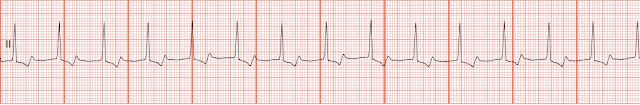
6.

a. Normal sinus rhythm
7.
 a. Sinus tachycardia with unifocal ventricular ectopy
8.
 a. Sinus tachycardia
9.

a. Sinus arrhythmia
10.

a. Atrial paced
Answers
1.
 c. Accelerated junctional rhythm. No P waves are present. The rate is 83. Based upon this information the rhythm fits the characteristics of an accelerated junctional rhythm
2.
 a. 3rd degree heart block. There is no relationship between the P waves and the QRS complexes. The P-P rhythm is regular and the R-R rhythm are both regular but at different rates.
3.
 c. AV pacing with multiform PVCs. The 6th and 8th complex (the first and 2nd PVC) appear to be a fusion complexes. This occurs when a PVC and a pacing stimulus occur simultaneously.
4.
 d. Sinus flutter with bigeminal PVCs. There is some baseline changes that make interpreting this strip somewhat difficult. Between the 2nd and 3rd complexes there are some flutter waves. The flutter waves are not as apparent between the 3rd and 4th complexes but reoccur elsewhere. The PVCs are unifocal in nature and occur every other beat.
5.
 c. Sinus rhythm with frequent unifocal PVCs
6.
 a. Normal sinus rhythm. Though not as dramatic as a lethal rhythm, a little normalcy in life is always good.
7.
 b. Atrial fibrillation with multifocal PVCs. The rhythm is irregular. The are no identifiable P waves present. The QRS complex is very wide with and RSR appearance. Probably a LBBB since it is positive monphasic wave in lead II. The ejection fraction on this patient cannot be very good.
8.
 c. Junctional tachycardia. The P waves are absent.
9.

d. Ventricular fibrillation. The patient is pulseless and apenic. CPR- defibrillation 200 J biphasic (360J monophasic)- CPR- Epinephrine (or Vasopressin)- Analyze rhythm- Defibrillation 300 J biphasic (360 monophasic)...
10.

b. Ventricular paced. A small pacer spike is noted before each QRS complex.
Reviewed 6/4/13
1.
b. Idioventricular
rhythm
c. Accelerated junctional
rhythm
d. Atrial
fibrillation with slow ventricular response
2.
a. 3rd degree heart block
b. Atrial flutter
with slow ventricular response
c. Bradycardia with sinus
arrhythmia
d. 2nd degree
heart block type II
3.
a. Atrial paced with unifocal PVCs
b. Ventricular
paced with atrial ectopic beats
c. AV pacing with multiform PVCs
d. Biventricular
pacing with PJCs
4.
b. Atrial fibrillation with multifocal PACs
c. Sinus
arrhythmia with frequent multifocal PACs
d. Sinus flutter with bigeminal PVCs
5.
b. Normal sinus
rhythm with a couplets of PVCs
c. Sinus rhythm
with frequent unifocal PVCs
d. Sinus rhythm
with premature junctional contractions
6.
a. Normal sinus rhythm
b. Sinus
bradycardia
c. Sinus
tachycardia
d. 1st degree AV
block
7.
b. Atrial fibrillation with multifocal PVCs
c. Sinus arrhythmia with frequent multifocal PACs
d. Sinus flutter with bigeminal PVCs
8.
b. Atrial flutter with rapid ventricular response
c. Junctional tachycardia
d. Supraventricular tachycardia
9.
b. Atrial fibrillation
c. Torsades de pointe
d. Ventricular fibrillation
10.
a. Atrial paced
b. Ventricular
paced
c. AV pacing
d. Biventricular
pacing
Answers
1.
2.
3.
4.
5.
6.
7.
8.
9.
d. Ventricular fibrillation. The patient is pulseless and apenic. CPR- defibrillation 200 J biphasic (360J monophasic)- CPR- Epinephrine (or Vasopressin)- Analyze rhythm- Defibrillation 300 J biphasic (360 monophasic)...
10.
b. Ventricular paced. A small pacer spike is noted before each QRS complex.
Reviewed 6/4/13


Comments
Post a Comment