Posts

Showing posts from October, 2016

Biventricular Pacing

Image
Biventricular pacing

Sinus Rhythm with Runs of Paroxysmal Atrial Tachycardia

Image
Sinus rhythm with runs of paroxysmal atrial tachycardia.  Notice the change in the morphology of the P waves.

Long Run of Ventricular Tachycardia

Image
These two consecutive pages on the same patient show a long run of VT.

Sinus Rhythm Changing to SVT

Image
Sinus Rhythm Changing to SVT

Sinus Bradycardia with a Dropped Beat and Sinus Srrest

Image
Sinus bradycardia with a dropped beat and sinus arrest

Atrial fibrillation with a Short Run of VT

Image
Atrial fibrillation with a short run of VT

Acute MI

Image
Acute MI: ST elevation, consider anterolateral injury or acute infarct ST elevation, consider inferior injury or acute infarct Should we consider a posterior wall MI?

Wide Complex Tachycardia Changing to VT.

Image
Wide Complex Tachycardia Changing to VT.    These three consecutive pages are on the same patient.  The patient had an underlying wide QRS complex.  You can see a transition in the morphology of the QRS complexes.  This was enough to suggest VT.      

Wide Complex Tachycardia

Image
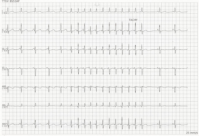
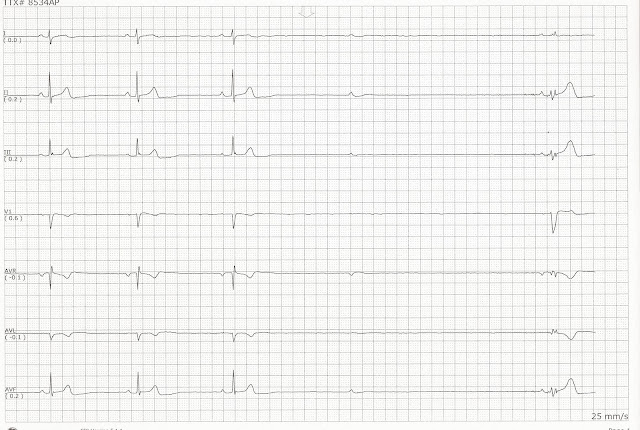
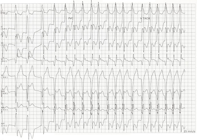
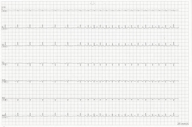
Wide Complex Tachycardia.  The pages are on the same pages.  The first page shows the underlying sinus rhythm so you can evaluate the QRS complexes.  The second page shows the same patient with a faster heart rate, around 150 bpm  The morphology of the QRS complexes is the same as the underlying rhythm seen on the first page.  Without knowing the underlying rhythm it would be easy to confuse the fast rhythm for VT.  

Atrial flutter

Image
Atrial flutter  

Idioventricular rhythm

Image
 Idioventricular rhythm  

Wolf Parkinson White

Image
Wolf-Parkinson-White  

Atrial Fibrillation with Ventricular Escape Beats

Image
Atrial Fibrillation with Ventricular Escape Beats.  These are two consecutive pages on the same patient.  She was in an atrial fibrillation with slow ventricular response.   She began having some runs of this ventricular escape rhythm.   

Run of Ventricular tachycardia

Image
Run of Ventricular tachycardia.  These are consecutive pages of a patient with a long run of VT.  It begins with what looks like an accelerated idioventricular rhythm which progresses to VT.  The rhythm spontaneously resolves and he returns to sinus rhythm.    

Atrial fibrillation with periods of arrest

Image
Atrial fibrillation with periods of arrest  

Wolf Parkinson White

Image
Wolf Parkinson White

AV Paced with a Run of VT

Image
AV Paced with a Run of VT  

Sinus Rhythm with a Run of Ventricular Tachycardia

Image
Sinus Rhythm with a Run of Ventricular Tachycardia  

Sinus Rhythm with a Run of Ventricular Tachycardia

Image
Sinus Rhythm with a Run of Ventricular Tachycardia.  These two consecutive pages are from the same patient who had a long run of VT  

Sinus Bradycardia with a 4 Beat Run of VT

Image
Sinus bradycardia with a 4 beat run of VT  

Ventricular paced with a run of VT

Image
Ventricular paced with a run of VT:  Complexes 1-5 are ventricular paced.  These are followed by an 8 beat run of VT.  A pause follows the run of VT and an fusion beat is seen.  The last two complexes are native or intrinsic beats.  

First degree block with a run of polymorphic VT

Image
Sinus rhythm with first degree block with a run of polymorphic VT.  Complexes 1 - 6 show a first degree block.  Complex 7 is an isolated PVC.  This is followed by a paced fusion beat.  During the run of VT another paced fusion beat is seen.   

Sinus rhythm with a run of VT

Image
Sinus rhythm with a run of VT:  The two pages are a continuation of the same run of VT    

Sinus rhythm changing to SVT

Image
Sinus rhythm changing to SVT  

Sinus bradycardia changing to second degree heart block type II

Image
Sinus bradycardia changing to second degree heart block type II  

Junctional rhythm.

Image
Junctional rhythm.