Posts

Showing posts from December, 2014

Practice EKG Strips 378

Image
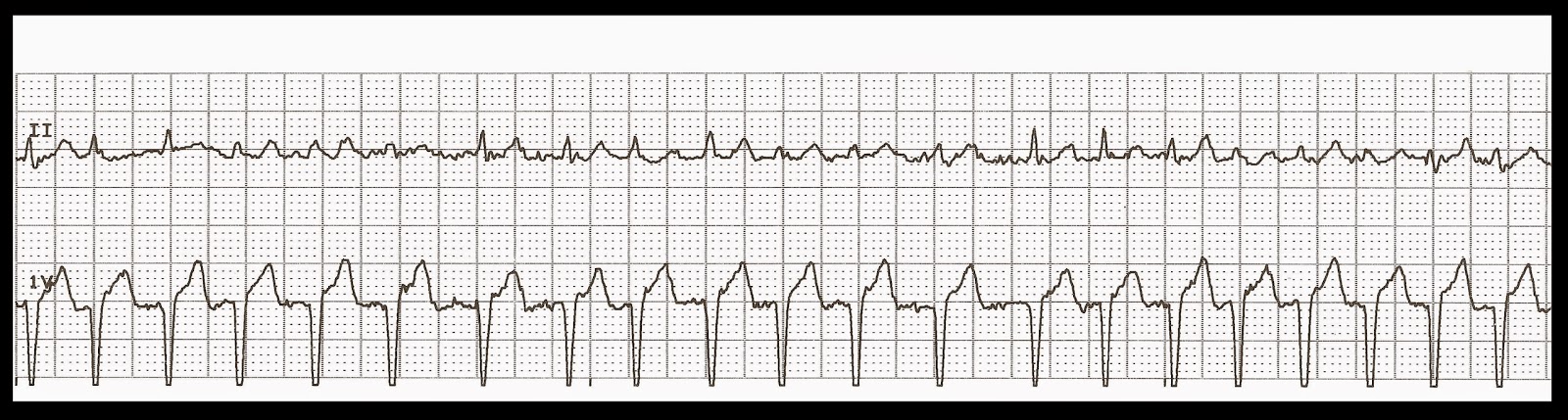
Identify the following rhythms. 1. a. Sinus tachycardia b. Ventricular tachycardia c. Atrial fibrillation with RVR d. Supraventricular tachycardia 2. a. Atrial paced b. Ventricular paced c. Dual paced d. Biventricular paced 3. a. Normal sinus rhythm b. First degree block c. Sinus bradycardia d. Second degree heart block type II 4. a. NSR with sinus arrest and an atrial escape beat b. NSR with sinus arrest and an junctional escape beat c. NSR with sinus arrest and an ventricular escape beat d. NSR with sinus arrest and an supraventricular escape beat 5. a. Polymorphic VT b. Coarse VF c. MAT d. SVT with aberrancy Answers 1. c. Atrial fibrillation with RVR. The rhythm is irregular with a rate of 140 bpm. No P waves can be readily identified. Some fibrillation is seen, especially in the V1 lead, between the QRS complexes. If the patient is symptomatic, consider rate slowing medications such as

Practice EKG Strips 377

Image
Identify the following rhythms. 1. a. Sinus arrhythmia b. Sinus rhythm with PACs c. Sinus rhythm with PVCs d. Sinus rhythm with pauses 2. a. Idioventricular rhythm b. Ventricular tachycardia c. Junctional rhythm d. Complete heart block 3. a. Atrial fibrillation b. Ventricular fibrillation c. Torsades de pointes d. Ventricular tachycardia 4. a. Demand atrial pacing b. Demand AV pacing c. Demand biventricular pacing d. Demand ventricular pacing 5. a. 2nd degree heart block type I b. 2nd degree heart block type II c. 1st degree heart block d. Sinus bradycardia with dropped PACs Answers 1. b. Sinus rhythm with PACs. The rhythm is irregular due to the frequent PACs. The heart rate is around 70 bpm. The PACs are the 3rd, 6th, and 8th complexes. The P waves of complexes 3 and 6 are fused with the T waves of the preceding complexes. A small P wave is seen with the 8th complex. 2.  a. Idioventricula

Practice EKG Strips 376

Image
Identify the following rhythms. 1. a. Atrial fibrillation b. Atrial flutter c. Atrial tachycardia d. Atrial paced 2. a. NSR with a triplet of PVCs b. Sinus arrhythmia with a triplet of PVCs c. First degree block with a triplet of PVCs d. Accelerated junctional rhythm with a triplet of PVCs 3. a. Sinus arrhythmia b. Normal sinus rhythm c. Sinus rhythm with a pause d. Sinus tachycardia 4. a. Normal sinus rhythm with sinus arrest b. Sinus bradycardia with a pause c. Sinus arrhythmia, sinus bradycardia d. Complete heart block 5. a. Atrial flutter changing to VT b. Sinus rhythm changing to VT c. Sinus tachycardia changing to VT d. Accelerated junctional changing to VT Answers 1. b. Atrial flutter. The rhythm is irregular with a rate of 90 bpm. No P waves are seen just some inverted or retrograde flutter waves. Atrial flutter is thought to be due to a re-entry type mechanism. The con

Practice EKG Strips Merry Christmas

Image
Identify the following rhythms. 1. a. NSR with PJCs b. NSR with PACs c. NSR with PVCs d. Sinus rhythm with pauses 2. a. NSR with sinus arrest b. 2nd degree heart block type II c. Sinus arrhythmia d. NSR with dropped PACs 3. a. Accelerated idioventricular rhythm b. Complete heart block c. Ventricular paced d. Accelerated junctional rhythm 4. a. Atrial flutter b. Atrial fibrillation c. Atrial tachycardia d. Atrial paced 5. a. Complete heart block b. Junctional rhythm c. Sinus arrhythmia d. Sinus bradycardia with a pause 6. a. Idioventricular rhythm with a PVC b. Complete heart block with a PVC c. Junctional rhythm with a PVC d. Sinus bradycardia with a PVC 7. a. Junctional rhythm b. Atrial fibrillation c. Sinus bradycardia d. Complete heart block 8. a. Supraventricular tachycardia b. Ventricular tachycardia c. Multifocal atrial tachycardia d

Practice EKG Strips 374

Image
Identify the following rhythms. 1. a. Atrial paced b. AV paced c. Biventricular paced d. Ventricular paced 2. a. Atrial paced with a PAC b. AV paced with a PAC c. Biventricular paced with a PAC d. Ventricular paced with a PAC 3. a. Demand atrial pacing with frequent PVCs b. Demand AV pacing with frequent PVCs c. Demand biventricular pacing with frequent PVCs d. Demand ventricular pacing with frequent PVCs 4. a. Atrial paced b. AV paced c. Biventricular paced d. Ventricular paced 5. a. Atrial paced b. AV paced c. Biventricular paced d. Ventricular paced Answers 1. a. Atrial paced. The rhythm is regular with a rate of 60 bpm. Very small P waves can be seen in lead V1 after the atrial pacer spike. The QRS complex is wide, .16 sec. It looks like an RSR complex lead V1 which is consistent with a RBBB. There is T wave inversion in the inferior lead II. A very long QT interval is seen of .5

Practice EKG Strips 373

Image
Identify the following rhythms. 1. a. Atrial fibrillation with slow ventricular response b. Sinus bradycardia with sinus arrhythmia c. Complete heart block d. Junctional rhythm 2. a. Atrial fibrillation b. Sinus tachycardia c. Atrial flutter d. SVT 3. a. Atrial paced with a couplet b. Ventricular paced with a couplet c. Dual paced with a couplet d. Biventricular paced with a couplet 4. a. NSR with a PJC b. NSR with a PAC c. NSR with PVC d. NSR with supraventricular beat 5. a. 2nd degree heart block type I b. 2nd degree heart block type II c. First degree block d. Complete heart block Answers 1. a. Atrial fibrillation with slow ventricular response. The rhythm is irregular. The rate is around 40 bpm (The last complex falls outside the 6 second marker). No P waves are seen just some fibrillation. No ectopic beats are seen. The QRS complex is narrow. 2.  c. Atrial flutter with a little

Practice EKG Strips 372

Image
Identify the following rhythms. 1. a. Normal sinus rhythm with a run of SVT b. Normal sinus rhythm with a run of VT c. Normal sinus rhythm with a run of AIVR d. Normal sinus rhythm with a run of ST 2. a. Junctional rhythm b. Complete heart block c. Sinus bradycardia d. Idioventricular rhythm 3. a. Sinus bradycardia with ST depression and a PVC b. Sinus rhythm with ST depression and a PVC c. Junctional rhythm with ST depression and a PVC d. First degree block with ST depression and a PVC 4. a. Ventricular tachycardia b. Atrial fibrillation c. Ventricular fibrillation d. Ventricular standstill 5. a. NSR with a PVC followed by more PVCs b. NSR with a PVC followed by junctional escape beats c. NSR with a PVC followed by atrial escape beats d. NSR with a PVC followed by some PACs Answers 1.  a. Normal sinus rhythm with a run of SVT. The run is fairly regular and there is an absence of P

Practice EKG Strips 371

Image
Identify the following rhythms. 1. a. Sinus tachycardia b. Supraventricular tachycardia c. Atrial fibrillation with RVR d. Ventricular tachycardia 2. a. Complete heart block b. Junctional rhythm c. Sinus bradycardia d. Sinus arrhythmia 3. a. Sinus tachycardia b. Sinus rhythm c. Accelerated junctional rhythm d. Accelerated idioventricular rhythm 4. a. Atrial paced with a couplet b. AV paced with a couplet c. Biventricular paced with a couplet d. Ventricular paced with a couplet 5. a. NSR with multifocal PVCs b. NSR with bigeminal PVCs c. NSR with trigeminal PVCs d. NSR with quadrigeminal PVCs Answers 1. c. Atrial fibrillation with RVR. Overall I think it is afib with RVR. At times it looks like there might be some P waves present but they are not consistent. If it were MAT then we would see more consistent P wave of varying morphology. If it were SVT then we would see a very regular rhythm. I

Practice EKG Strips 370

Image
Identify the following rhythms. 1. a. Atrial paced b. Biventricular paced c. Dual paced d. Ventricular paced 2. a. Normal sinus rhythm changing to VT b. Sinus bradycardia changing to VT c. Atrial fibrillation changing to VT d. Idioventricular rhythm changing to VT 3. a. Accelerated idioventricular rhythm b. Accelerated junctional rhythm c. Idioventricular rhythm d. Junctional rhythm 4. a. NSR with a sinus exit block b. NSR with a PAC c. Sinus arrhythmia d. Sinus rhythm with a dropped PAC 5. a. Sinus arrhythmia b. Sinus rhythm with sinus arrest c. Sinus rhythm with bigeminal PACs d. 2nd degree heart block type II Answers 1. d. Ventricular paced. A single pacer spike is seen just before the QRS complex. 2. c. Atrial fibrillation changing to VT. The initial rhythm is irregular with an absence of P waves. This changes into a ventricular tachycardia with a rate between 150 - 180 bpm. 3. b. Acce