Posts

Showing posts from December, 2012

Code Blue: PEA

Image
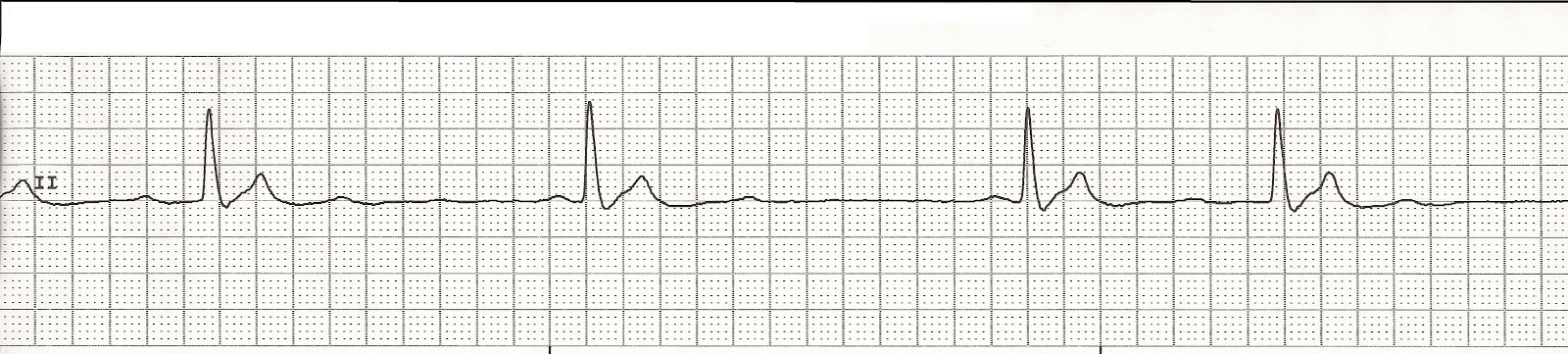
Near, far, wherever you are  I believe that the heart does go on…  09:53  Placed on monitor Initial rhythm 09:55 CPR Unsuccessful IV access attempt Unsuccessful IV access attempt CPR Rhythm 10:00 CPR Intubated 7.5 ET tube.  Tube placement confirmed CPR Rhythm 10:02 Epinephrine 2mg via ET tube Successful IO placement in right tibia CPR in progress CPR Rhythm 10:05 Vasopressin 40 units IO CPR in progress CPR Rhythm Check 10:09 Epinephrine 1mg IO NS fluid bolus started CPR Rhythm 10:11 Sodium Bicarbonate IO CPR in progress Adequate ventilations CPR Rhythm Check 10:15 Epinephrine 1mg IO CPR in progress CPR Rhythm 10:19 Epinephrine 1mg IO CPR in progress CPR Rhythm Check 10:24 Epinephrine 1mg IO CPR in progress CPR Rhythm 10:27  Epinephrine 1mg IO Continued CPR Final rhythm 10:28  Code ended Once more you open the door And you're here

Pediatric PEA Part 5

Pediatric PEA E valuate Reversible Causes ·          Hypoxia ·          Hypovolemia ·          Hyper/hypokalemia ·          Hypothermia ·          Tension pneumothorax ·          Tamponade ·          Thromboembolism ·          Tables/toxins ·          Trauma Note:   Management of PEA is exactly the same as for asystole. Interventions for specific causes of PEA may lead to successful resuscitation. Appropriate measures may include a rapid fluid bolus for potential hypovolemia (20 mL/kg normal saline or lactated Ringer’s administered IV or IO), needle decompression for suspected tension pneumothorax, ventilation and sodium bicarbonate for acidosis, or pericardiocentesis for cardiac tamponade as regional protocols permit. Reversible Causes of PEA ·          Hypoxia- use DOPE mnemonic ·          Hypovolemia- 20ml/kg fluid bolus ·          Hyper/hypokalemia- correct imbalance ·          Hypothermia- consider rewarming techniques Note:   DOPE mnemoni

Pediatric PEA Part 4

Image
Pediatric PEA Length based color-coded tape. Medication Dose Calculation ·          Use the child’s weight if it is known ·          If the child’s weight is unknown, it is reasonable to use a body length tape ·          No data regarding the safety or efficacy of adjusting the doses for obese patients IV Access ·          Peripheral IV ·          Central line ·          Intraosseous ·           Endo tratracheal Peripheral IVs ·           Placement may be difficult in a critically ill child ·           Central venous placement requires procedure can be time consuming Central IV Drug Delivery •                      Peak drug concentrations are higher and drug circulation times shorter •                      Central line placement can interrupt CPR. •                      A central line extending into the superior vena cava can be used to monitor ScvO2 and estimate CPP during CPR, both of which are predictive of ROSC Intraosseous (IO)

Pediatric PEA Part 3

Image
Pediatric PEA Secondary Survey ·          Intubate ·          Oxygenate ·          IV access ·          Treat reversible causes Note: Once the patient is intubated, continue CPR with asynchronous ventilations and chest compressions. Formula for Estimating Endotracheal tube size:  Uncuffed ET tube:  mm ID = (age in years/4) + 4 Cuffed ET tube:  mm ID = (age in years/4) + 3.5 Confirm ET tube placement n   Direct cord visualization n   End-tidal CO2 monitor n   Purple- problem n   Yellow- yes n   Tan- think about it n   Bilateral breath sounds n   CXR n   Continuous waveform capnography Note:  Continuous quantitative waveform capnography is now recommended for intubated patients throughout the periarrest period as a means of both confirming and monitoring correct placement of an endotracheal tube.  Esophageal Detector Device (EDD) ·          May be considered in children weighing ≥ 20 kg with a perfusing rhythm ·          Insufficient

Pediatric PEA part 2

Image
Pediatric PEA Rhythms Idioventricular rhythm Sinus bradycardia Sinus tachycardia with inverted T waves Agonal Rhythm PEA mnemonic:  Keep them ALIVE A ssess ABCs L isten for pulse using doppler I nitiate CPR V asoconstrictors E valuate reversable causes A ssess ABCs- Primary Survey ·          Assess responsiveness and pulse ·          Active EMS system ·          Call for defibrillator/monitor Note:  If a rhythm is present on the monitor but the pulse is absent (eg,PEA), CPR should be started immediately, beginning with chest compressions, and should continue for 2 minutes before the rhythm check is repeated. L isten for Pulse Using Doppler ·          A doppler will help distinguish between a pulseless state and profoundly weak cardiac contractions with a low cardiac output (pseudo-PEA). ·          True PEA:  no pulse and no perfusion ·          Pseudo-PEA: weak pulse detected by doppler or echocardiography and sev

Pediatric PEA Part 1

Pediatric PEA Pediatric Non-profusing Rhythms ·          Pulseless electrical activity ·          Asystole ·          Agonal Rhythms ·          Ventricular tachycardia ·          Ventricular fibrillation Cardiac arrest in infants and children ·          Does not usually result from a primary cardiac cause ·          Terminal result of progressive respiratory failure or shock ·          Asphyxia begins ·          Period of systemic hypoxemia, hypercapnia, and acidosis ·          Progresses to bradycardia and hypotension ·          Culminates with cardiac arrest Pulseless Electrical Activity ·          Displays a rhythm on the monitor but does not have an arterial pulse ·          Confirm pulselessness with doppler ·          Key to treatment is CPR and early identification of possible causes ·          Outcome is very poor unless the cause can be established and treated ·          Pediatric patients who are in PEA do not benefit from defibri

Pediatric cardiac arrest: PEA

Image
1250:  A 10 year old boy is brought to the emergency room by EMS after he struck a tree with his three-wheeler.  On the scene he was conscious,  alert and oriented but in route he began to have increasing shortness of breath and was becoming more lethargic.  The EMTs initiated positive pressure ventilations in route to the hospital.    He is on a spinal board with a C collar in place.   When he is transferred from the EMS stretcher to the hospital stretcher is found to be unresponsive and he no longer has spontaneous respirations.  1252:  No pulse is detected.  A pediatric code is called and CPR is initiated.  He is placed on the monitor and this is his initial rhythm.  PEA rhythm 1255: CPR is continued.   A #20 IV is started in his right antecubtial fossa.  Epinephrine 1mg IV is given and and a fluid bolus of 20ml/kg is also started.  Because there is difficulty with providing positive pressure ventilations, the patient is intubated with a 6.5 cuffed ET tube. 

EKG Rhythm Strips 89

Image
Identify the following rhythms 1. 2. 3. 4. 5. Answers 1.  Atrial fibrillation with couplets The rhythm is irregular with a rate of 80/min.   No P waves are present but fibrillatory activity can be seen betweeen the QRS complexes.  There are multifocal PVCs present, some in couplets.  PR: ---,  QRS:  .10 sec,  QT:  .36 sec. 2.  Bradycardia with 1st degree block. The rate is 56/min.   The rhythm is regular.   There are upright, uniform P waves before each QRS complex.  No ectopic beats are present.  PR:  0.28 sec,  QRS:  .08 sec,  QT:  .42 sec. 3.  NSR with PACs. The rhythm is irregular due to atrial ectopy.   The rate is  90/min.   There are upright P waves befoe each QRS complex.  Also, there is a small nonconducted P wave after the first PAC.   Premature atrial complexes are present.  PR:  .16 sec,  QRS:  .08 sec,  QT:  .40 sec. 4.  NSR with unifocal PVCs (trigeminy) The rhythm is irregular due to the ectopic beats.   The rate is