Posts

Showing posts from September, 2016

Sinus bradycardia changing to a ventricular escape rhythm

Image
Sinus bradycardia changing to a ventricular escape rhythm  

Failure to Capture Part II

Image
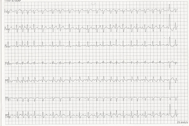
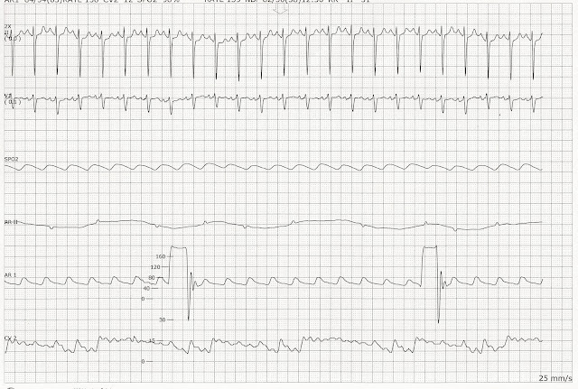
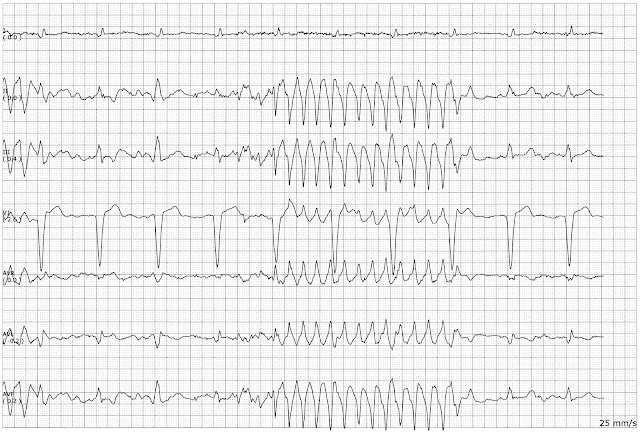
The patient was becoming cyanotic.  He did not have a palpable pulse.  By the time we could get BP machine to the room, the episode had passed.  He was noted to have his cell phone lying over his ICD/pacemaker so there is a question that the episode may have been induced by the cell phone.  The pacemaker was interrogated.   

Failure to Capture Part 1

Image
The patient complained of "not feeling" well and shortness of breath.   

Sinus rhythm with a run of ventricular tachycardia

Image
Sinus rhythm with a run of ventricular tachycardia  

Wide complex tachycardia changing to sinus bradycardia

Image
Wide complex tachycardia changing to sinus bradycardia  

2nd degree heart block type I

Image
2nd degree heart block type I  

Sinus rhythm with dropped PACs

Image
Sinus rhythm with dropped PACs  

Sinus rhythm with a run of ventricular tachycardia

Image
Sinus rhythm with a run of ventricular tachycardia  

Sinus bradycardia with unifocal PVCs and nonconducted P wave

Image
Sinus bradycardia with unifocal PVCs and nonconducted P wave  

MVP Mode Pacemaker

Image
We recently had a patient with an AV pacemaker that appeared to have periods of failure to pace. The pacemaker representative evaluated the rhythm and said that the pacemaker appeared to be set in the "MVP" mode.  The MVP mode works in the manner describe in this diagram      

Sinus rhythm with non-sustained VT

Image
Sinus rhythm with a non-sustained run of VT.          

Atrial fibrillation with slow ventricular response

Image
Atrial fibrillation with slow ventricular response.  In the V1 lead it looks more like an atrial flutter.  The conduction through the AV node is variable and slow.   

Second degree heart block type II

Image
Second degree heart block type II  
Image
Atrial flutter.   

Sinus bradycardia with an AV dissociation

Image
  Sinus bradycardia with an AV dissociation.  Notice the shortening PR interval on the 3rd - 6th complexes.              

Junctional escape beats

Image
A dropped PAC is seen after the 2nd complex.  This is followed by three junctional escape beats.  A PAC occurs (6th complex) and another junctional escape beat is seen (7th complex)  

Complete heart block.

Image
Complete heart block.  The rhythm is irregular.  The QRS complexes are narrow too.  This suggests that the block is higher in the ventricle.  The rate is faster too.   

Multifocal atrial tachycardia

Image
Multifocal atrial tachycardia.  There are at least 3 different looking P waves throughout the strip.  A fusion beat is seen on the 2nd to the last complex.   

Sinus rhythm changing to accelerated idioventricular rhythm

Image
Sinus rhythm changing to accelerated idioventricular rhythm.  In lead II there appear to be some retrograde P waves that are following the QRS complexes.   

Ventricular paced with some fusion complexes

Image
Ventricular paced with some fusion complexes.  The 3rd, 9th, and 10th complexes are the fusion complexes.   

Paroxysmal atrial tachycardia

Image
Paroxysmal atrial tachycardia.  Notice the change in the morphology of the P waves as the rhythm transitions back into a sinus rhythm.  

Arterial blood pressure waveform

Image
Sinus tachycardia.  This patient had an arterial line and a CVP.  The square wave test was done on the arterial line to evaluate the system dynamics.  Two to three oscillations should follow a fast flush of the system.   

Quadrigeminal PVCs

Image
Quadrigeminal PVCs.  Unifocal PVCs are seen every fourth beat.  

Sinus rhythm changing to accelerated idioventricular rhythm

Image
Sinus rhythm changing to accelerated idioventricular rhythm (AIVR).  The second page is a continuation of the first page.   

Trigeminal PVCs

Image
Sinus rhythm with trigeminal PVCs.   

Movement artifact

Image
Movement artifact.  The omplexes are organized with artifact in between the complexes in leads I and V1.  

Polymorphic ventricular tachycardia

Image
Polymorphic ventricular tachycardia  

Wolf-Parkinson-White

Image
Wolf-Parkinson-White