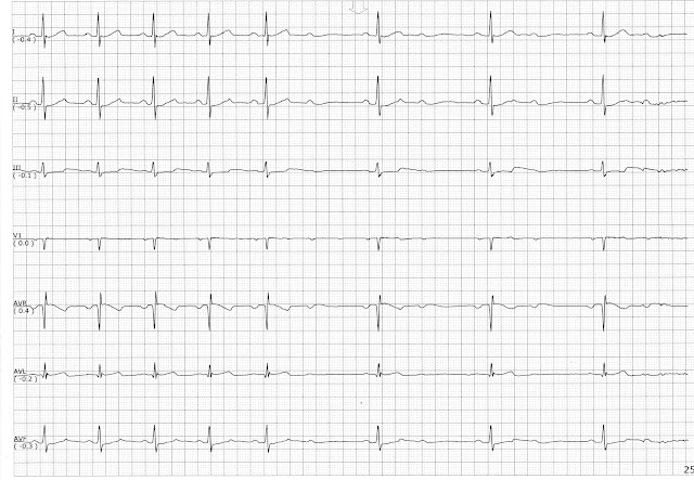
Posts

Showing posts from March, 2016

A Dropped PAC

Image
On this page a dropped PAC is seen following the 5th complex.  If an impulse is initiated during the phase of relative refractory period an impulse may or may not produce a contraction.  Generally, it takes a stronger than normal impulse to initiate a contraction.

2nd Degree Heart Block Type I

Image
On this page you can see the progressive prolongation of the PR interval over successive beats.  The dropped beat is not seen in lead I but you can see it clearly in leads II and aVR.  A PVC interrupts the cycle of beats.  Instead of continuing in the cycle, the cycle of beats starts over after a compensatory pause.

Overdrive Pacing

Image
On this page the pacemaker is either attempting to override the fast heart rate or accelerating to keep up with the patient's physical demands.  Any thoughts?

Atrial fibrillation or MAT

Image
At first glance I would have called this atrial fibrillation with RVR but upon closer exam, there appears to be some P waves of varying morphology present.

Atrial fibrillation with a Run of Ventricular Tachycardia

Image
This page shows a patient in atrial fibrillation who had a nonsustained run of VT.  The patient converted out of the VT without any intervention.

Atrial flutter

Image
This page show a patient with atrial flutter with a long period of arrest.

Ventricular Tachycardia Converting to Sinus Rhythm

Image
This patient had a run of non-sustained ventricular tachycardia that spontaneously converted to sinus rhythm.

Atrial Fibrillation with Slow Ventricular Response

Image
On this page the page has atrial fibrillation with slow ventricular response.  We are more accustomed to seeing atrial fibrillation with a rapid ventricular response.  However, when the patient takes too much medication or has an electrolyte problem we can see a slower atrial fibrillation.

Extreme Bradycardia with Sinus Arrest

Image
On this page the rhythm shows extreme sinus bradycardia.  The patient had a history of sick sinus syndrome.  

Intermittent Type I Block

Image
This patient had an intermittent type I block.  He would have periods of sinus rhythm and then have periods of 2nd degree type I block.  The grouping on the type I block was not always consistent.  It may have been 3 beats in one group and 5 beats in another group.

Fusion beats and Nonconducted PACs

Image
On this page there are a variety of complexes present.  The first complex shows a prolonged PR interval.  The 2nd and 3rd complexes are PVCs with P waves or fusion complexes.  On the 3rd complex there is a dropped P wave.  It can be seen in lead aVR and aVL.  This is followed by a short pause.  The 4th complex shows a prolonged PR interval and it is followed by another nonconducted P wave.  A short pause follows the nonconducted P wave, then sinus bradycardia with sinus arrhythmia follows. 

Sinus Rhythm Changing to VT or ST with Aberrancy

Image
On this page the rhythm starts out with sinus but changes to a wide complex tachycardia.  At the beginning of the wide complex tachycardia there are P waves associated with the QRS complexes so this seems to suggest some kind of aberrancy rather than VT.  The axis of the QRS complexes does not change during the accelerated rhythm either.

Sinus Rhythm Changing to AIVR

Image
This page also shows a sinus rhythm changing to an accelerated idioventricular rhythm.  Notice the very short PR interval on the initial complexes of the AIVR.  The QRS complexes look like they have a delta wave configuration too.

Sinus Rhythm Changing to AIVR

Image
This page shows a patient in sinus rhythm that converts to an accelerated idioventricular rhythm.

Biventricular Pacing with Trigeminal PVCs

Image
This rhythm shows biventricular pacing with trigeminal PVCs.  The third PVC is also a multifocal triplet.  The PVCs have a pacer spike fused in them.  

2nd Degree Heart Block Type II

Image
This patient was having intermittent episodes of a 2nd degree type II block. 

Overdrive pacing

Image
This patient was in a wide complex tachycardia.   There appears to be an underlying atrial fibrillation. His pacemaker successfully overrides the rhythm and a ventricular paced rhythm follows the fast rhythm.

Run of SVT

Image
This rhythm page begins with sinus rhythm.  The second complex appears to be a PAC and it is followed by a brief run of supraventricular tachycardia.  The morphology of the QRS complexes in lead III change during the run of SVT but overall the morphology and QRS complex width remains the same.  Sinus rhythm resumes afterwards. 

Accelerated Idioventricular rhythm

Image
This patient was initially in sinus rhythm but converted to an accelerated idioventricular rhythm.

Ventricular Tachycardia with a Significant Drop in the Arterial Blood Pressure

Image
On this page, the patient has a ventricular pacemaker.  He has a long run of VT with a significant drop in his arterial blood pressure.  This strip shows why a person can become hemodynamically unstable with VT.

Ventricular paced with a Brief Run of VT

Image
On this page, the patient is ventricular paced and has a non-sustained run of VT.  On the bottom rhythm you can see a brief drop in the arterial blood pressure associated with the VT.

Ventricular Tachycardia Changing to Bigeminy

Image
In this strip, the patient is in AIVR or VT and converts to atrial fibrillation with bigeminal PVCs

Ventricular Bigeminy

Image
On this page, the patient is in atrial fibrillation and is having some bigeminal PVCs