Posts

Showing posts from April, 2013

Basic EKG Rhythm Test 07

Image
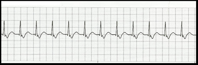
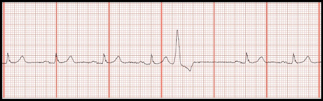
Identify the following rhythms. 1. 2. 3. 4. 5. 6. 7. 8. 9. 10. 11. 12. 13. 14. 15. 16. 17. 18. 19. 20. 21. 22. 23. 24. 25. Do you want to try another Basic EKG Rhythm Test? Click here. Answers 1.    Junctional tachycardia 2.    Accelerated junctional rhythm 3.    Atrial paced 4.    Normal sinus rhythm with PACs 5.    Sinus rhythm changing to ventricular tachycardia 6.    Supraventricular tachycardia 7.    Ventricular tachycardia changing to ventricular fibrillation 8.    2nd degree heart block type I 9.    Atrial fibrillation with multifocal PVCs 10.  Idioventricular rhythm 11.  Normal sinus rhythm with a multifocal couplet 12.  Polymorphic ventricular tachycardia 13.  

Practice EKG Rhythm Strips 126

Image
Identify the following rhythms. 1. 2. 3. 4. 5. Answers 1. Normal sinus rhythm with a PVC The underlying rhythm is regular.   The calculated rate is 65.   There are upright P waves that precede the QRS complexes.   There is a single ectopic PVC.   PR:  .20 sec.   QRS:  .10 sec.  QT:  .40 sec 2. Normal sinus rhythm with a PJC The underlying rhythm is regular.   The calculated rate is 62.    There are upright P waves before each sinus QRS complex.   A single PJC is noted, the 3rd complex.   The P wave of this complex is retrograde in origin.   PR:  12 sec,  QRS:  .08 sec,  QT:  .44 sec. 3. Normal sinus rhythm 1st degree block with a pause The rhythm is irregular due to the pause.   The rate is 80 per minute (10 sec strip rule).   There are upright P waves before each QRS complex.  Notice that the P waves are wide, about .12 sec.  There may be some underlying left atrial enlargement.   No ect

Practice EKG Rhythm Strips 125

Image
Identify the following rhythms. 1. 2. 3. 4. 5. Answers 1.  Agonal rhythm.   The QRS complex is wide and bizzare looking owing to it's ventricular origin.   The rate is less than 20 minute so this qualifies this rhythm as an agonal rhythm. 2. Atrial paced.  The P waves that follow the atrial paced spike are not very prominent.   There is a significant delay between the atrial spike and the QRS complex.  This is supposed to help reduce the development of a pacemaker induced hypertrophy.   Otherwise, the rhythm is regular.   The rate is 75.  There are upright P waves that are atrial paced in origin.   No ectopy is noted.   PR:  32 sec.   QRS:  .08 sec,   QT:  .32 sec. 3. Junctional rhythm.  .   Those T waves are tall so I wonder if there was a problem with hyperkalemia.  The rhythm is regular.   The rate is 60.   There are no P waves before each QRS complex.  No ectopic beats are noted.   PR: ---   QRS:  .12 sec,  QT:  

Practice EKG Rhythm Strips 124

Image
Identify the following rhythms. 1. 2. 3. 4. 5. Answers 1. Sinus tachycardia with a wandering baseline The rhythm looks regular, though this is difficult to tell because of the wandering baseline.   The calculated rate is 150/min.   There are upright P waves before each QRS complex.  The P waves are tall and wide suggesting biatrial enlargement.   No ectopy is noted.   PR:  12 sec.   QRS.  08 sec,   QT:  .28 sec. 2. Normal sinus rhythm with sinus pause The rhythm is irregular owing to the pause, otherwise the underlying rhythm is regular.   The calculated rate is 60/minute.   There are upright P waves before each QRS complex.   There is a pause lasts 1.4 sec.  PR:  .20 sec,  QRS:  .08 sec,  QT:  .38 sec. 3. Sinus arrhythmia The rhythm is irregular.   Notice how the R-R interval slowly gets longer.   The R-R interval between the 1st and 2nd complex is .6 sec.   The R-R interval betw