Posts

Showing posts from June, 2013

Basic EKG Rhythm Test 09

Image
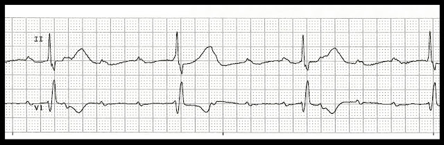
Identify the following rhythms 1. 2. 3. 4. 5. 6. 7. 8. 9. 10. 11. 12. 13. 14. 15. 16. 17. 18. 19. 20. 21. 22. 23. 24. 25. Do you want to try another Basic EKG Rhythm Test? Click here Answers 01.    Atrial fibrillation 02.    Sinus tachycardia 03.    Ventricular paced 04.    Accelerated junctional rhythm 05.    Sinus bradycardia with bigeminal PVCs 06.    Junctional rhythm 07.    Multifocal atrial tachycardia 08.    1st degree AV block with PJCs 09.    2nd degree AV block type I 10.    Normal sinus rhythm 11.    Agonal rhythm

Practice EKG Rhythm Strips 155

Image
Identify the following rhythms. 1. 2. 3. 4. 5. Answers 1. There is AV dissociation.  The atrial rate is regular with a rate of 107/min and a ventricular rate of 32/min.  The P waves are positive and uniform but have no relationship to a QRS complex.  No ectopic beats are present.  PR:  ---,  QRS:  .16 sec,  QT:  .66 sec. 2. The rhythm is irregular.  The rate is 70/min.  The P waves are splint but are associated with a QRS complex.  The 3rd complex occurs early and has a change in the P wave morphology.   This is especially evident in the lower V1 lead.  This is followed by a junctional escape beat, 4th complex,  then a sinus beat.   The 6th complex is another PAC.   This is followed by another sinus looking beat.  PR:  .20 sec,  QRS:  .10 sec,  QT:  .40 sec. 3. Junctional rhythm The rhythm is regular with a rate of 50/min.  Retrograde P waves precede the QRS complex.  No ectopic beats are pr

Practice EKG Rhythm Strips 154

Image
Identify the following rhythms. 1. 2. 3. 4. 5. Answers 1. Spraventricular tacycardia The rhythm is regular.   The rate is 166/min.  No readily identifiable P waves are present.  No ectopic beats are present.  The QRS is narrow, 0.08 sec.  The QT interval is 0.28 sec. 2. Sinus tachycardia with 1st degree block and a PVC The rhythm is irregular due to the PVC.  The rate is 110/min.  The P waves are upright and precede QRS complex.  The PR interval is prolonged.  A single PVC is present and it coupled with a P wave.  PR:  .24 sec,  QRS:  .08 sec,  QT:  .36 sec. 3. 1st degree block changing to 2nd degree type II The rhythm starts out as a 1st degree block with a PR interval of .32 and then transitions into a type I block and then a type II block. 4. Polymorphic ventricular tachycardia changing to sinus bradycardia The strip begins with polymorphic ventricular tachycardia which abruptly change

Practice EKG Rhythm Strips 153

Image
Identify the following rhythms. 1. 2. 3. 4. 5. Answers 1. Agonal rhythm Agonal rhythm is a slow idioventricular rhythm with a rate of less than 20.   A terminal rhythm that often precedes asystole and death. 2. Demand pacing with unifocal PVCs The rhythm is irregular due to the ectopic beats.   The rate is  60/min.   The are some native P waves as well as some that are pacemaker generated.  On the first complex the P wave is generated by the patient.  The PR interval is prolonged and measures .26 sec.  On the subsequent complexes a P wave is not sensed by the pacemaker so the atrial pacemaker generates an impulse within it's set parameters.  The ventricle is completely paced.  Two unifocal PVCs are present.  PR:  .24 sec, QRS:  .16 sec,  QT:  .48 sec. 3. Sinus pause The rhythm is irregular because of the pause.  The rate is 60/min.  The P waves are upright and precede the QRS complex but the PR interval is prolonged.