Posts

Showing posts from June, 2014

Basic EKG Rhythm Test 20

Image
Identify the following rhythms. 01. a. First Degree block b. Second degree block type I c. Second degree block type II d. Third degree block 02. a. Atrial paced b. Biventricular paced c. Dual paced d. Ventricular paced 03. a. Sinus bradycardia b. Junctional rhythm c. Idioventricular rhythm d. Agonal rhythm 04. a. Atrial flutter b. Atrial fibrillation c. 2nd degree heart block type II d. NSR with PACs 05. a. Atrial paced with a multifocal couplet b. Biventricular paced  with a multifocal couplet c. Dual paced  with a multifocal couplet d. Ventricular paced  with a multifocal couplet 06. a. Polymorphic ventricular tachycardia b. Ventricular fibrillation c. Torsades de pointes d. Atrial fibrillation 07. a. NSR with a pause b. Sinus arrhythmia c. Wandering atrial pacemaker d. Atrial fibrillation 08. a. NSR with bigeminal PVCs b.

Practice Rhythm Strips 306

Image
Identify the following rhythms. 1. a. Asystole b. Ventricular paced c. Atrial paced d. Biventricular paced 2. a. NSR with couplets b. NSR with ventricular bigeminy c. NSR with uniform PVCs d. NSR with unifocal PACs 3. a. Accelerated idioventricular rhythm b. Accelerated junctional rhythm c. Complete heart block d. Agonal rhythm 4. a. 1st degree block with a triplet of PVCs b. Sinus rhythm with triplet of PVCs c. Sinus bradycardia with triplet of PVCs d. Sinus rhythm with trigeminal PVCs 5. a. Sinus bradycardia with a pause b. Sinus arrhythmia c. Complete heart block d. 2nd degree AV block type II Answers 1. a. Asystole .   Asystole with a functioning single chamber pacemaker. 2. b. NSR with ventricular bigeminy.  The rhythm is irregular because of the PVCs. The heart rate is 90/min. The P waves look the same and are paired with a QRS complex. The QRS complexes are narrow. Unifocal PVCs are seen i

Practice Rhythm Strips 305

Image
Identify the following rhythms. 1. a. Junctional rhythm b. Sinus bradycardia c. Complete heart block d. Idioventricular rhythm 2. What is the rhythm?  a. Ventricular tachycardia  b. Multifocal atrial tachycardia  c. Sinus tachycardia  d. Supraventricular tachycardia 3. a. Complete heart block b. Junctional rhythm c. Idioventricular rhythm d. Ventricular paced 4. a. 2nd degree AV block type II b. Atrial flutter c. Sinus tachycardia d. 2nd degree AV block type I 5. a. NSR with sinus arrest b. Complete heart block c. Junctional rhythm d. Sinus bradycardia Answers 1. b. Sinus bradycardia.  The rhythm is regular and the rate is 50/min. The P waves are paired with QRS complexes. The QRS complexes are narrow. No ectopic beats are seen. PR: .16 sec, QRS: .08 sec, QT: .44 sec. Interpretation: Sinus bradycardia 2. d. Supraventricular tachycardia.  The rhythm is regular with a rate of 214mn. No P waves are seen. Th

Practice Rhythm Strips 304

Image
Identify the following rhythms. 1. a. Ventricular fibrillation b. Ventricular tachycardia c. Asystole d. Ventricular standstill 2. a. NSR with PACs b. NSR with some PJCs c. NSR with a couple of PVC d. NSR with escape beats 3. a. 1st degree block b. Wenckebach c. Mobitz II d. Complete heart block 4. a. Sinus bradycardia with a multifocal couplet b. NSR with multifocal PVCs c. Sinus arrhythmia with uniform PVCs d. Sinus tachycardia with bigeminal PVCs 5. a. Ventricular standstill b. Asystole c. Agonal rhythm d. Sinus bradycardia Answers 1. a. Ventricular fibrillation.  Juice 'em up at 200 J. 2.  a. NSR with PACs 3.  b. Wenckebach .  Those P waves are drifting away from the QRS complexes. The rhythm is irregular with a rate of 50/min. The P waves are positive and uniform. There is progressive elongation of the PR interval over successive beats.  Interpretation:  2nd degree AV Block Type II or just

Practice Rhythm Strips 303

Image
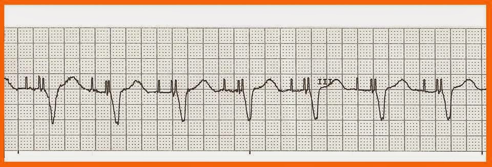
Identify the following rhythms. 1. a. Atrial paced b. Biventricular paced c. Dual paced d. Ventricular paced 2. a. Normal sinus rhythm with couplets b. Normal sinus rhythm with multifocal PVCs c. Normal sinus rhythm with multiform PVCs d. Normal sinus rhythm with bigeminal PVCs 3. a. Idioventricular rhythm b. Sinus bradycardia c. Junctional rhythm d. Agonal rhythm 4. a. Sinus bradycardia with 1st degree block b. Junctional rhythm with 1st degree block c. Idioventricular rhythm with 1st degree block d. 2nd degree AV block type I 5. a. 1st degree AV block b. 2nd degree AV block type I c. 2nd degree AV block type II d. 3rd degree block Answers 1.  c. Dual paced.  The rhythm is regular. The rate is 70/min. There are atrial pacer spikes seen before the P waves and Biventricular pacer spikes seen before each QRS complex. No ectopic beats are noted. PR: .20 sec, QRS: .16 sec, QT: .48 sec. Interpretation: D

Practice Rhythm Strips 302

Image
Identify the following rhythms. 1. a. Sinus tachycardia b. Ventricular tachycardia c Supraventricular tachycardia d. Multifocal atrial tachycardia 2. a. Atrial paced b. AV paced c. Biventricular paced d. Ventricular paced 3. a. 1st degree AV block b. Idioventricular rhythm c. Agonal rhythm d. Complete heart block 4. a. Torasades de pointe b. Monomorphic ventricular tachycardia c. Polymorphic ventricular tachycardia d. Multifocal atrial tachycardia 5. a. Normal sinus rhythm with multifocal PVCs b. Atrial fibrillation with multifocal PVCs c. Sinus arrhythmia with multifocal PVCs d. Sinus bradycardia with multifocal PVCs Answers 1. a. Sinus tachycardia .  There are inverted T waves present. So those deflections before the QRS complexes are probably atrial in origin.  The rhythm is regular with rate of 150/min. There are upright P waves that precede the QRS complexes. The QRS complexes are narrow. There are