Posts

Showing posts from August, 2013

Basic EKG Rhythm Test 11

Image
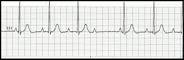
Identify the following rhythms. 1. 2. 3. 4. 5. 6. 7. 8. 9. 10. 11. 12. 13. 14. 15. 16. 17. 18. 19. 20. 21. 22. 23. 24. 25. 26. 27. Do you want to try another Basic EKG Rhythm Test? Then click here Answers 1.    Idioventricular rhythm 2.    2nd degree heart block type II 3.    Junctional tachycardia 4.    2nd degree heart block type I 5.    Agonal rhythm 6.    1st degree heart block 7.    Asystole with pacer spikes 8.    Atrial fibrillation 9.    Atrial flutter 10.  AV paced 11.  3rd degree heart block 12.  Idioventricular rhythm 13.  Junctional rhythm 14.  Accelerated junctional rhythm 15.  NSR with triplets of PVCs 16.  NSR with bigeminal PACs 17.  NSR with multifocal bigeminal PVCs 18.  NSR with trigeminal

Practice EKG Rhythm Strips 188

Image
Identify the following rhythms. 1. 2. 3. 4. 5. Answers 1. 2nd degree AV block type I The rhythm is irregular with a heart rate of 50/min.   The P waves are upright.   There is a nonconducted P wave after the 1st and 3rd complexes.  The PR interval gets progressively longer then this followed by a nonconducted P wave.   No ectopic beats are present.  PR:  .12/.24/.32 sec, QRS:  .08 sec,  QT:  .40 sec. 2. Idioventricular rhythm The rhythm is irregular with a rate of 30/min.  No P waves are present.  The QRS complex is very wide.  The rhythm is a ventricular escape rhythm usually seen in a dying heart.  It certainly looks agonal but based on the rate it is still called an idioventricular rhythm. 3. 3rd degree heart block The rhythm is regular with a ventricular rate of 40/min and an atrial rate of 90/min.   The P waves are dissociated from the QRS complexes.   No ectopic beats are noted.  

Practice EKG Rhythm Strips 187

Image
Identify the following rhythms. 1. 2. 3. 4. 5. Answers 1. Normal sinus rhythm changing to accelerated junctional rhythm The rhythm is regular with a rate of 88/min.   There are both upright and negative P waves present.   The P waves of the first four complexes are upright but beginning at the fifth complex the P waves are retrograde.   No ectopic beats are present.   PR:  .12 sec,  QRS:  .08 sec,  QT:  . 36 sec. 2. Seven beat run of ventricular tachycardia The rhythm is irregular.   The rate is 120/min.   Upright P waves are seen on the first 4 beats.  The 1st and 3rd beats are sinus beats while the 2nd and 4th beats look like PACs.   This is followed by a 7 beat run of ventricular tachycardia.   PR:  .12 sec,  QRS:  .08 sec,  QT:  .36 sec. 3. Bigeminal PACs The rhythm is irregular with frequent PACs.    The rate is 100/min.   There are upright P waves present.   The P waves are associat

Practice EKG Rhythm Strips 186

Image
Identify the following rhythms. 1. 2. 3. 4. 5. Answers 1. 2nd degree AV block type I The rhythm is irregular with a rate of 50/min.  The P-P interval is regular while the R-R interval is irregular.  The P waves are upright and uniform.   There is a nonconducted P wave that follows the 3rd complex.   No ectopic beats are noted.   PR:  .12/.24/.32 sec,  QRS:  .08 sec,  QT:  .40 sec. 2. Ventricular paced with multifocal couplets The rhythm is irregular due to the PVCs.   The paced rate is 88/min.   Upright P waves are paired with a QRS complex.   There are two multifocal couplets present.  PR:  .20 sec,  QRS:  .16 sec,  QT:  .40 sec. 3. 3rd degree heart block The rhythm is regular with a ventricular rate of 30/min and an atrial rate of 90/min.  The P waves are upright and uniform but are unassociated with a QRS complex.   PR:  ---,  QRS:  .16 sec,  QT: .40 sec. 4. Idioventricular rhyth