Posts

Showing posts from May, 2012

EKG Rhythm Strips 51

Image
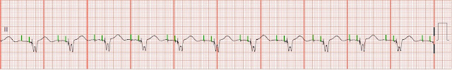
Identify the following rhythm strips. 1. 2. 3. 4. 5. Answers 1. Atrial fibrillation with unifocal PVCs The rhythm is irregular with some PVCs.   The rate is 100/min.    No P waves are present.   Some fibrillatory activity is seen between the QRS complexes.   It looks like there might be P waves present before the 5th complex and following but this appears to be part of the ST segment.  If you look at the 1st complex you will see that following the T wave there is a slight depression followed by a slight positive rise.   This appears to be repeated in the complexes that follows giving the impression that a P wave is present.   The increase in the heart rate and shortening of the R-R interval would also give the impression that P waves are present.   Two unifocal PVCs are seen.  PR:  ---,  QRS:  .12 sec,  QT:  .38 sec.   The bottom tracing is an arterial blood pressure waveform.   You can seen the hemodynamic effects of the PVCs on the arteria

EKG Rhythm Strips 50

Image
Identify the following rhythms. 1. 2. 3. 4. 5. Answers 1. Atrial flutter with variable ventricular response The rhythm is irregular with a rate of 80/min.   Flutter waves are seen between the QRS complexes.   The ratio of flutter waves to QRS complexes varies.   No ectopic beats are identified.   PR:  ---,  QRS:  .16 sec,  QT:  .40 sec 2. Atrial fibrillation with slow ventricular response The rhythm is irregular with a ventricular rate of 48/min.   P waves are absent but some fibrillation is noted between the QRS complexes.  No ectopic beats are seen.   PR:  ---,  QRS:  .12 sec,  QT:  .48 sec. 3. Atrial paced The rhythm is regular with a rate of 60/min.   Atrial pacer spikes precede the very small P waves.  No ectopic beats are noted.  PR:  .20 sec,  QRS:  .08 sec,  QT:  .44 sec. 4. Junctional rhythm The rhythm is regular.   The rate is 32/min.  The P waves are absent.   No e

EKG Rhythm Strips 49

Image
Identify the following rhythms. 1. 2. 3. 4. 5. Answers 1. Junctional tachycardia The rhythm is slightly irregular with a rate of 102/min.  The P waves are inverted and precede the QRS complex.   No ectopic beats are noted.   PR:  .16 sec,  QRS:  .08 sec,  QT:  .40 sec. 2. Atrial fibrillation with rapid ventricular response The rhythm is irregular.   The ventricular rate is 180/min.   No P waves are clearly seen.   There is some fibrillation noted between some of the complexes.    No ectopic beats are noted.  PR:  ---,  QRS:   .08 sec,  QT:  .26 sec. 3. Accelerated idioventricular rhythm The rhythm is regular.   The rate is 72/min.   No P waves are seen.   No ectopic beats are noted.  The QRS complex is wide.   The T waves are tall.   A wide slow rhythm with tall T waves suggests hyperkalemia.   PR:  ---,  QRS:  .20 sec,  QT:  .72 sec. 4. 1 st degree AV block with unifocal PVCs and a couplet of PVCs

EKG Rhythm Strips 48

Image
Identify the following rhythms 1.  2. 3. 4. 5. Answers 1. AV paced with biventricular pacing The rhythm is regular with a rate of 75/min.   Both atrial and biventricular pacer spikes are seen before the P waves and the QRS complexes.   PR:  .20 sec,  QRS:  .16 sec,  QT:  .48 sec. 2. Atrial paced The rhythm is regular.   The ventricular rate is 65/min.  An atrial pacing stimulus is seen before each P wave.  No ectopic beats are seen.  PR:  .20 sec,  QRS:  .08 sec,  QT:  .48 sec. 3. Atrial paced The rhythm is regular.   The heart rate is 60/min.  Atrial pacer spikes are seen before the P waves. No ectopic beats are seen.  The ST segment is isoelectric but the T wave is inverted.  PR:  .20 sec, QRS:  .08 sec,  QT:  .44 sec. 4. Ventricular paced The rhythm is regular with a ventricular rate of 68/min.  There are flutter/fibrillatory waves seen between the QRS complexes.   A ventricular pacing

An Unusual AV Dissociation

Image
At first glance this has the appearance of a 2nd degree type I AV block.   See if you can identify what is unusual about it. In a 2nd degree type I AV block (Wenckebach) the PR interval gets progressively longer until there is a non conducted P wave or dropped QRS complex.   In most cases the P-P interval is regular.  Often the block occurs at the level of the AV node and has a narrow QRS complex.   The rhythm is usually benign and requires no immediate treatment  In a 2nd degree type II block the conducted P waves have a consistent PR interval but there are some non-conducted P waves present.  The non conducted P waves may have a 2:1, 3:1 ratio.   The level of the block is around the bundle of HIS.    With complete heart block the atria and ventricles each have an independent rhythm.   The P waves are not associated with the QRS complexes.  If the level of the block is higher in the His-purkenje system then the QRS complex will have a narrow appearance with a junc

Megacode PEA Part 7

Tablets ·          Assess for drug overdose ·          Assess for hx of drug use ·          Consult poison control Tablets: Calcium Channel Blockers ·          Hypotension ·          Bradycardia with variable heart block ·          Altered mental status ·          EKG: slow rate, prolonged PRI, AV blocks ·          Rx Calcium chloride 10% 1-4G slow IV ·          Fluid resuscitation, vasopressor agents, atropine, transvenous pacing Tablets: Beta blockers ·          Hypotension ·          Bradycardia with variable heart block ·          EKG: slow rate, prolonged PRI ·          Rx Glucagon 3-10mg IV bolus followed by infusion 2-5mg/hr Tablets: Tricyclic Antidepressants ·          Amitriptyline, Doxepine, Trazadone, Nortriptyline ·          Prolonged QT, Widened QRS ·          3Cs & 1A - Cardiac dysrhythmias, Convulsions, Coma, & Acidosis ·          Rx NaHCO3 bolus or infusions ·          Maintain pH > 7.45 Tablets: Digoxin

Megacode PEA Part 6

Image
Hypovolemia ·          Look for tachycardia with narrow QRS complex ·          Assess and find bleeding sources ·          Fluid boluses with isotonic crystaloids ·          Volume expanders ·          Blood products Hydrogen Ions (acidosis) ·          Assess ABGs ·          Bicarb 1mEq/kg o    Known pre-existing bicarbonate responsive acidosis o    Intubated patient with continued long arrest interval o    Upon return of spontaneous circulation after long arrest interval o    Tricyclic antidepressant or aspirin overdose o    Known prexisting hyperkalemia ·          ·Treat respiratory acidosis with ventilation Hyperkalemia Assess serum potassium levels Tall T waves, widened QRS 3.5-5.3             Normal 5.3-6.0             Mild 6.1-7.0             Moderate >7.0                Severe Hypokalemia Hypothermia ·          Assess core temperature ·          Cardiac dysrrhythmias when <

Megacode PEA Part 5

Image
V asoconstrictors ·          Epinephrine ·          Vasopressin Note:  Available evidence suggests that the routine use of atropine during PEA or asystole is unlikely to have a therapeutic benefit. For this reason, atropine has been removed from the Cardiac Arrest Algorithm. To date no placebo-controlled trials have shown that administration of any vasopressor agent at any stage during management of VF, pulseless VT, PEA, or asystole increases the rate of neurologically intact survival to hospital discharge. There is evidence, however, that the use of vasopressor agents is associated with an increased rate of ROSC. Drug Therapy: Epinephrine ·          Epinephrine 1mg (1:10,000 solution) IV/IO q3-5min ·          ETT dose 2mg diluted in 10cc of NS ·          Increases systemic vascular resistance (vasoconstriction) ·          Increase coronary and cerebral perfusion pressures during CPR ·          Escalating or high doses without demonstrable benefit ·     

Megacode PEA Part 4

Image
Peripheral IV Drug Delivery ·          Adults peak drug concentrations are lower and circulation times longer ·          Does not require interruption of CPR ·          Administer the drug by bolus injection and follow with a 20-mL bolus of IV fluid ·          Elevate the extremity for 10 to 20 seconds to facilitate drug delivery to the central circulation. Endotracheal Drug Delivery ·          Results in lower blood concentrations than the same dose given intravascularly. ·          Give 2 to 2½ times the recommended IV dose. ·          Providers should dilute the recommended dose in 5 to 10 ml of water or normal saline and inject the drug directly into the endotracheal tube Intraosseous Drug Delivery ·          Enables drug delivery similar to that achieved by peripheral venous access at comparable doses. ·          Is safe and effective for fluid resuscitation, drug delivery, and  blood sampling ·          Is attainable in all age groups.