EKG Rhythm Strips 51
Identify the following rhythm strips.
1.

2.
 3.
3.
 4.
4.
 5.
5.

Answers
1.
The rhythm is irregular with some PVCs. The rate is 100/min. No P waves are present. Some fibrillatory activity is seen between the QRS complexes. It looks like there might be P waves present before the 5th complex and following but this appears to be part of the ST segment. If you look at the 1st complex you will see that following the T wave there is a slight depression followed by a slight positive rise. This appears to be repeated in the complexes that follows giving the impression that a P wave is present. The increase in the heart rate and shortening of the R-R interval would also give the impression that P waves are present. Two unifocal PVCs are seen. PR: ---, QRS: .12 sec, QT: .38 sec. The bottom tracing is an arterial blood pressure waveform. You can seen the hemodynamic effects of the PVCs on the arterial pressure and pulse waveform.
2.
The rhythm is irregular due to the demand pacing. The rate is 80/min. Both sinus and atrial paced P waves are present. The 5th and 6th complexes have sinus P waves. Two multifocal PVCs are present. The ventricles are completely paced. PR: .14 sec, QRS: .16 sec, QT: .44 sec.
3.
The rhythm is irregular with a rate of 70/min. The P waves are upright and have a corresponding QRS complex. Two multifocal PVCs are present. A compensatory pause follows the PVCs. PR: .20 sec, QRS: .08 sec, QT: .44 sec.
4.
The rhythm is regular. The rate is 43/min. The P waves are uniform, upright, and associated with a QRS complex. No ectopic beats are noted. PR: .16 sec, QRS: .08 sec, QT: .48 sec.
5.
The rhythm is irregular with a rate of 132/min. P waves are present and are paired with a QRS complex. There are PACs that closely follow the 7th and 20th complexes. The P wave of the PAC is hidden in the T wave of the previous complex. Notice the change in the height of the T wave. PR: .16 sec, QRS: .16 sec, QT: .32 sec.
Reviewed 3/2/16
1.

2.


Answers
1.
|
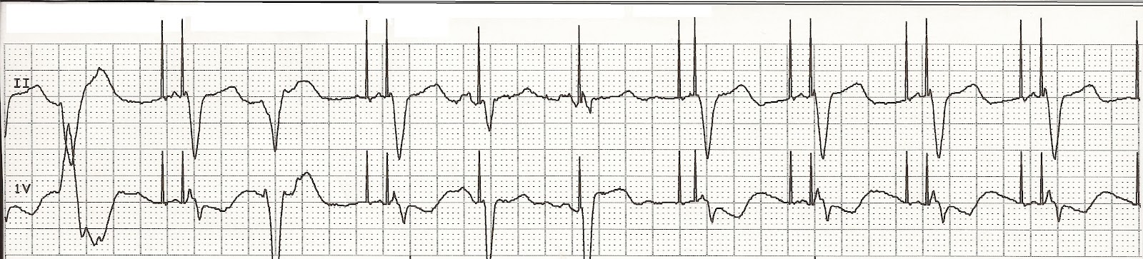
| Atrial fibrillation with unifocal PVCs |
The rhythm is irregular with some PVCs. The rate is 100/min. No P waves are present. Some fibrillatory activity is seen between the QRS complexes. It looks like there might be P waves present before the 5th complex and following but this appears to be part of the ST segment. If you look at the 1st complex you will see that following the T wave there is a slight depression followed by a slight positive rise. This appears to be repeated in the complexes that follows giving the impression that a P wave is present. The increase in the heart rate and shortening of the R-R interval would also give the impression that P waves are present. Two unifocal PVCs are seen. PR: ---, QRS: .12 sec, QT: .38 sec. The bottom tracing is an arterial blood pressure waveform. You can seen the hemodynamic effects of the PVCs on the arterial pressure and pulse waveform.
2.
|
| Demand atrial pacing with multifocal PVCs |
The rhythm is irregular due to the demand pacing. The rate is 80/min. Both sinus and atrial paced P waves are present. The 5th and 6th complexes have sinus P waves. Two multifocal PVCs are present. The ventricles are completely paced. PR: .14 sec, QRS: .16 sec, QT: .44 sec.
3.
|
| Normal sinus rhythm with multifocal PVCs |
The rhythm is irregular with a rate of 70/min. The P waves are upright and have a corresponding QRS complex. Two multifocal PVCs are present. A compensatory pause follows the PVCs. PR: .20 sec, QRS: .08 sec, QT: .44 sec.
4.
|
| Sinus bradycardia |
The rhythm is regular. The rate is 43/min. The P waves are uniform, upright, and associated with a QRS complex. No ectopic beats are noted. PR: .16 sec, QRS: .08 sec, QT: .48 sec.
5.
|
| Sinus tachycardia with PACs |
The rhythm is irregular with a rate of 132/min. P waves are present and are paired with a QRS complex. There are PACs that closely follow the 7th and 20th complexes. The P wave of the PAC is hidden in the T wave of the previous complex. Notice the change in the height of the T wave. PR: .16 sec, QRS: .16 sec, QT: .32 sec.
Reviewed 3/2/16


Comments
Post a Comment