EKG Rhythm Strip Quiz 34
Identify the following rhythms.
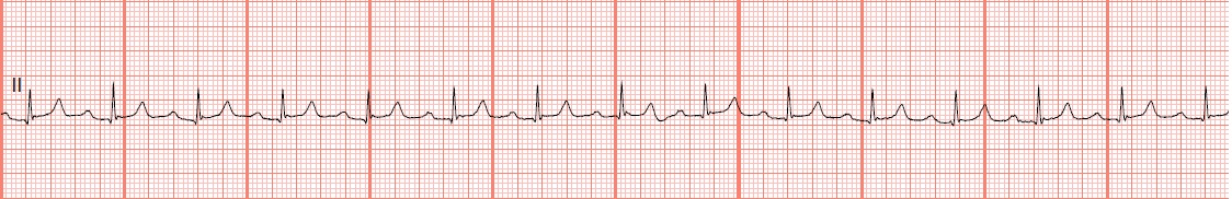
a. Normal sinus rhythm. My favorite rhythm at the end of a long day. Good night!
Reviewed 6/4/13, 3/2/16
1.
.JPG)
a. Sinus bradycardia with atrial ectopy
2.
.jpg)
a. Sinus bradycardia with atrial ectopy
3.
 a. Normal sinus rhythm
4.
 a. Sinus tachycardia
5.
 a. Atrial paced
6.
 a. Normal sinus rhythm with 1st degree block
7.
 a. Complete heart block
.JPG)
a. Sinus bradycardia with atrial ectopy
b. Normal sinus
rhythm with unifocal PVCs
c. Sinus
tachycardia with frequent multifocal PACs
d. Sinus rhythm
with premature junctional contractions
2.
.jpg)
a. Sinus bradycardia with atrial ectopy
b. Normal sinus
rhythm with unifocal PVCs
c. Sinus
tachycardia with frequent multifocal PACs
d. Sinus rhythm
with premature junctional contractions
3.

b. Sinus
bradycardia
c. Sinus
tachycardia
d. Sinus bradycardia with 1st degree AV block4.

b. Atrial flutter with rapid ventricular response
c. Multifocal atrial tachycardia
d. Supraventricular tachycardia
5.

b. Ventricular
paced
c. AV pacing
d. Biventricular
pacing
b. Sinus
bradycardia
c. Sinus
tachycardia
d. 2nd
degree heart block type II
7.

b. Normal sinus rhythm with 1st degree block
c. Accelerated junctional rhythm
d. Accelerated idioventricular rhythm
8.
 a. Atrial paced
9.
 a. Sinus bradycardia with atrial ectopy
b. Normal sinus rhythm with an occasional PAC
10.

1.
 d. Sinus rhythm with premature junctional contractions. The inverted P waves before each premature beat are characteristic of PJCs
2.
 b. Normal sinus rhythm with unifocal PVCs
3.
d. Sinus bradycardia with 1st degree AV block. The PR interval is .28. The P waves are split. Split P waves are sometimes referred to as P Mitrale and are seen in left atrial enlargement and mitral valve disease.
4.
d. Supraventricular tachycardia. The rate is around 150. No P waves are evident.
5.
b. Ventricular paced. Pacer spikes are seen before each QRS complex.
6.
a. Normal sinus rhythm with 1st degree block. The PR interval is 0.28 seconds.
7.
c. Accelerated junctional rhythm. Best guess, accelerated idioventricular rhythm with a prolonged QT interval of 0.60 seconds. The EKG machine called this an "undetermined rhythm." It could be sinus rhythm with a wide QRS complex and a very long 1st degree AV block (0.36 sec). The P wave could be wide due to left atrial enlargement. If this is the case then, as the saying goes (An American saying from the Apollo space program) "Houston we have a problem." It could be an accelerated junctional rhythm with a prolonged QT interval. You would have to know the patient's history to determine if the patient had a pre-existing bundle branch block, in lead II probably a LBBB. The EKG machine may be right and it is an undetermined rhythm. The bottom line, is treat the patient and not the machine. Please feel free to send me your comments.
8.
c. AV pacing. It looks like the underlying rhythm may be an atrial fibrillation. A pacer spike is seen before the P wave and the QRS complex.
9.
b. Normal sinus rhythm with an occasional PAC. The P wave is very small in comparison the the sinus P waves and has a shorter PR interval.
10.
c. Accelerated junctional rhythm
d. Accelerated idioventricular rhythm
8.

b. Ventricular
paced
c. AV pacing
d. Biventricular
pacing
9.

b. Normal sinus rhythm with an occasional PAC
c. Sinus
tachycardia with frequent multifocal PVCs
10.

a. Normal sinus rhythm
b. Sinus
bradycardia
c. Sinus
tachycardia
d. 1st degree AV
block
Answers

2.

3.
4.
d. Supraventricular tachycardia. The rate is around 150. No P waves are evident.
5.
6.
7.
c. Accelerated junctional rhythm. Best guess, accelerated idioventricular rhythm with a prolonged QT interval of 0.60 seconds. The EKG machine called this an "undetermined rhythm." It could be sinus rhythm with a wide QRS complex and a very long 1st degree AV block (0.36 sec). The P wave could be wide due to left atrial enlargement. If this is the case then, as the saying goes (An American saying from the Apollo space program) "Houston we have a problem." It could be an accelerated junctional rhythm with a prolonged QT interval. You would have to know the patient's history to determine if the patient had a pre-existing bundle branch block, in lead II probably a LBBB. The EKG machine may be right and it is an undetermined rhythm. The bottom line, is treat the patient and not the machine. Please feel free to send me your comments.
8.
9.
10.
Reviewed 6/4/13, 3/2/16


Thank you so much FloatRN-Mike for this blog!
ReplyDeleteMy favorite for EKG reviewing.
Thanks for all the quizzes and answers.
Thanks a lot!