Practice EKG Strips 399
Identify the following rhythms.
1.

a. Supraventricular tachycardia
b. Atrial fibrillation with RVR
c. Ventricular tachycardia
d. Multifocal atrial tachycardia
2.

a. Atrial flutter
b. 2nd degree heart block type II
c. Atrial fibrillation
d. 2nd degree heart block type I
3.

a. NSR with unifocal PACs
b. NSR with multifocal PVCs
c. NSR with bigeminal PVCs
d. NSR with multiform PVCs
4.

a. Sinus bradycardia with PACs
b. Sinus arrhythmia with PVCs
c. Complete heart block
d. Atrial fibrillation with slow ventricular
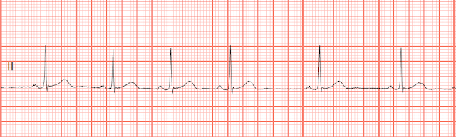
5.

a. Sinus bradycardia with a U wave
b. 2nd degree heart block type II
c. 2nd degree heart block type I
d. Sinus bradycardia with dropped PACs
Answers
1. Atrial fibrillation with RVR. The rhythm is very irregular so this cannot be SVT which has a regular rhythm. The QRS complexes appear to be a little wide but there appears to be some fibrillation between some of the QRS complexes. This is not something that you would see with VT. With MAT you would need to see some consistent P waves across the strip. On complexes 4, 5, and seven you can't be sure if that small positive deflection is part of the T wave. There appears to be some fibrillaton between the 7 - 8 and 11 - 12 complexes.
2. a. Atrial flutter. The rhythm is regular with a rate of 50 bpm. 3:1 flutter waves are seen between the QRS complexes. The QRS complexes are wide, .12 sec. No ectopic beats are seen.
3. c. NSR with bigeminal PVCs. The rhythm is irregular due to the PVCs. There upright P waves associated with the sinus QRS complexes. Unifocal PVCs every other beat. PR: .20 sec, QRS: .08 sec, QT: .40 sec.
4. c. Complete heart block. There is no relationship between the P waves and the QRS complexes. The P waves can be seen landing on the T waves and on the QRS complexes.
5. Sinus bradycardia with a U wave. The P - P interval is shorter from the P wave to what I will call the U wave when compared to the P - P interval from the U wave to the P wave. With a type II block the P - P interval is usually very regular. It would be very unusually to see so many consecutive dropped PACs, it is possible but nothing you would typically see.
1.
a. Supraventricular tachycardia
b. Atrial fibrillation with RVR
c. Ventricular tachycardia
d. Multifocal atrial tachycardia
2.
a. Atrial flutter
b. 2nd degree heart block type II
c. Atrial fibrillation
d. 2nd degree heart block type I
3.
a. NSR with unifocal PACs
b. NSR with multifocal PVCs
c. NSR with bigeminal PVCs
d. NSR with multiform PVCs
4.
a. Sinus bradycardia with PACs
b. Sinus arrhythmia with PVCs
c. Complete heart block
d. Atrial fibrillation with slow ventricular
5.
a. Sinus bradycardia with a U wave
b. 2nd degree heart block type II
c. 2nd degree heart block type I
d. Sinus bradycardia with dropped PACs
Answers
1. Atrial fibrillation with RVR. The rhythm is very irregular so this cannot be SVT which has a regular rhythm. The QRS complexes appear to be a little wide but there appears to be some fibrillation between some of the QRS complexes. This is not something that you would see with VT. With MAT you would need to see some consistent P waves across the strip. On complexes 4, 5, and seven you can't be sure if that small positive deflection is part of the T wave. There appears to be some fibrillaton between the 7 - 8 and 11 - 12 complexes.
2. a. Atrial flutter. The rhythm is regular with a rate of 50 bpm. 3:1 flutter waves are seen between the QRS complexes. The QRS complexes are wide, .12 sec. No ectopic beats are seen.
3. c. NSR with bigeminal PVCs. The rhythm is irregular due to the PVCs. There upright P waves associated with the sinus QRS complexes. Unifocal PVCs every other beat. PR: .20 sec, QRS: .08 sec, QT: .40 sec.
4. c. Complete heart block. There is no relationship between the P waves and the QRS complexes. The P waves can be seen landing on the T waves and on the QRS complexes.
5. Sinus bradycardia with a U wave. The P - P interval is shorter from the P wave to what I will call the U wave when compared to the P - P interval from the U wave to the P wave. With a type II block the P - P interval is usually very regular. It would be very unusually to see so many consecutive dropped PACs, it is possible but nothing you would typically see.


Comments
Post a Comment