Practice EKG Rhythm Strips 161
Identify the following rhythms.
1.

2.

3.

4.

5.

Answers
1.
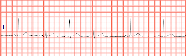
The rhythm appears regular though it is difficult to tell because of the wandering baseline. The rate is 70/min. There are upright P waves before each QRS complex. The P waves are split and suggest left atrial enlargement. No ectopy is noted. PR: .20 sec, QRS: .08 sec, QT: .40 sec. Sometimes lead II can be affected by the patient's respiratory pattern. For convenience sake, the upper limb leads are placed on the chest wall near the shoulder and the leg leads are placed on the abdomen. To correct the wandering baseline, move the arm leads further down the arm and the the leg leads further down the leg. This should eliminate any interference from the respiratory muscles or the abdomen if the patient is using accessory muscles to breath.
2.
The rhythm is irregular. The R-R interval between the 1st and 2nd complexes is .76 sec and the R-R interval between the 5th and 6th complexes is 1.12 sec. The P waves are upright and precede the QRS complexes. No ectopic beats are present. PR: .24 sec, QRS: .08 sec, QT: .40 sec
3.
The rhythm is regular. The rate is 115/min. The P waves are upright and precede the QRS complexes. The P waves are split suggesting left atrial enlargement. No ectopic beats are present. PR: .22 sec, QRS: .08 sec, QT: .30 sec.
4.
The rhythm is regular and the rate is 187/min. Small upright P waves are present and precede the QRS complex. No ectopic beats are noted. PR: .06 sec, QRS: .06 sec, QT: .24 sec
5.
The rhythm is irregular due to the unifocal PVCs. The rate is 80/min. The P waves are not associated with each QRS complex. There appears to be an underlying complete heart block. A P wave is seen in the ST segment of the 1st complex and another P wave is seen on the pacer spike of the 3rd complex. Two unifocal PVCs are present. There are ventricular pacer spikes present. PR: ---, QRS: .16 sec, QT: .44 sec.
1.
2.
3.
4.
5.
Answers
1.
|
| Normal sinus rhythm with wandering baseline |
The rhythm appears regular though it is difficult to tell because of the wandering baseline. The rate is 70/min. There are upright P waves before each QRS complex. The P waves are split and suggest left atrial enlargement. No ectopy is noted. PR: .20 sec, QRS: .08 sec, QT: .40 sec. Sometimes lead II can be affected by the patient's respiratory pattern. For convenience sake, the upper limb leads are placed on the chest wall near the shoulder and the leg leads are placed on the abdomen. To correct the wandering baseline, move the arm leads further down the arm and the the leg leads further down the leg. This should eliminate any interference from the respiratory muscles or the abdomen if the patient is using accessory muscles to breath.
2.
|
| Sinus arrhythmia with 1st degree block |
The rhythm is irregular. The R-R interval between the 1st and 2nd complexes is .76 sec and the R-R interval between the 5th and 6th complexes is 1.12 sec. The P waves are upright and precede the QRS complexes. No ectopic beats are present. PR: .24 sec, QRS: .08 sec, QT: .40 sec
3.
|
| Sinus tachycardia with 1st degree block |
The rhythm is regular. The rate is 115/min. The P waves are upright and precede the QRS complexes. The P waves are split suggesting left atrial enlargement. No ectopic beats are present. PR: .22 sec, QRS: .08 sec, QT: .30 sec.
4.
|
| Sinus tachycardia |
The rhythm is regular and the rate is 187/min. Small upright P waves are present and precede the QRS complex. No ectopic beats are noted. PR: .06 sec, QRS: .06 sec, QT: .24 sec
5.
|
| Ventricular paced with unifocal PVCs |
The rhythm is irregular due to the unifocal PVCs. The rate is 80/min. The P waves are not associated with each QRS complex. There appears to be an underlying complete heart block. A P wave is seen in the ST segment of the 1st complex and another P wave is seen on the pacer spike of the 3rd complex. Two unifocal PVCs are present. There are ventricular pacer spikes present. PR: ---, QRS: .16 sec, QT: .44 sec.


Comments
Post a Comment