Posts

Showing posts from February, 2012

Pediatric Advance Life Support: Asystole Part 4

Image
Medication Dose Calculation ·          Use the child’s weight if it is known ·          If the child’s weight is unknown, it is reasonable to use a body length tape ·          No data regarding the safety or efficacy of adjusting the doses for obese patients Broweslow Tape IV Access ·          Peripheral IV ·          Central line ·          Intraosseous ·          Intratracheal Intraosseous (IO) Access ·          All intravenous medications can be administered intraosseously ·          Onset of action and drug levels are comparable to venous administration ·          IO access can be used...

Pediatric Advance Life Support: Aystole Part 3

Confirm ET tube placement n   Direct cord visualization n   End-tidal CO2 monitor n   Purple- problem n   Yellow- yes n   Tan- think about it n   Bilateral breath sounds n   CXR n   Continuous waveform capnography Note:  Continuous quantitative waveform capnography is now recommended for intubated patients throughout the periarrest period as a means of both confirming and monitoring correct placement of an endotracheal tube.  Esophageal Detector Device (EDD) ·          May be considered in children weighing ≥ 20 kg with a perfusing rhythm ·          Insufficient data to recommend for or against its use in children during cardiac arrest Verification of Endotracheal Tube Placement ·          Verify proper tube placement immediately after intubation ·          After securing the ...

Pediatric Advance Life Support: Aystole Part 2

Image
Mnemonic for Treating Asystole:  CAVE C - CPR A -assess rhythm in another lead V -vasoconstrictors E - evaluate reversible causes CPR :  Primary Survey ·          Assess patient ·          Support ABCs ·          CPR ·          Attach defibrillator/monitor ·          Assess rhythm The effectiveness of PALS is dependent on high-quality CPR ·          Adequate compression rate (at least 100 compressions/min) ·          Adequate compression depth (at least one third of the AP diameter of the chest or approximately ·          1 1⁄2 inches [4 cm] in infants and approximately 2inches [5 cm] in children) ·          Allowing com...

Pediatric Advance Life Support: Asystole Part 1

Image
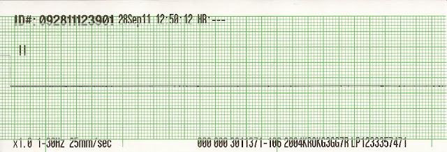
Non-profusing Rhythms ·          Pulseless electrical activity ·          Asystole ·          Agonal Rhythms ·          Ventricular tachycardia ·          Ventricular fibrillation Cardiac Arrest in Infants and Children ·          Does not usually result from a primary cardiac cause ·          Terminal result of progressive respiratory failure or shock ·          Asphyxia begins ·          Period of systemic hypoxemia, hypercapnia, and acidosis ·          Progresses to bradycardia and hypotension ·          Culminates with cardiac arrest Keys to Treati...

EKG Rhythm Strips 37

Image
Identify the following rhythms 1. 2. 3. 4. 5. Answers 1. NSR with multiple ectopic beats The rhythm is irregular with multiple ectopic beats.   The ventricular rate is 96/min. (Count number of R waves in this 10 strip and multiple by 6).  The P waves are upright and have a corresponding QRS complex.    The are multiple ectopic beats present, 2nd 6th, 10th, 12th, 13th and 15th complexes.   There appears to be a P wave associated with the 1st and 2nd ectopic beats these are actually be fusion beats.   The 12th and 13th complexes are PVCs that form a multifocal couplet.  The 15th complex is a PVC that is followed by a compensatory pause and an atrial escape beat.  PR:  .20 sec,  QRS;  .10 sec,  QT:  .40 sec. 2. NSR with unifocal PVCs The rhythm is irregular with multiple PVCs.   The P waves are positive and are associated with a QRS complex.   There are unifocal P...

EKG Rhythm Strips 36

Image
Identify the following rhythms 1. 2. 3. 4. 5. Answers 1. NSR with unifocal PVCs The rhythm is irregular with a heart rate of 78/min.    The P waves are upright, uniform and associated with a QRS complex.   Two unifocal PVCs are seen.   PR:  .12 sec,  QRS:  .08 sec, QT:  .44 sec. 2. Sinus bradycardia with a U wave The rhythm is regular with a rate of 44/min.  The P waves are uniform and are paired with a QRS complex.  The P waves are wide and slightly notched suggesting biatrial enlargement.  No ectopic beats are noted.  A U wave follows each T wave.PR:  .16 sec,  QRS:  .10 sec,  QT:  .56 sec.   3. Atrial fibrillation with rapid ventricular response.   The rhythm is irregular with a ventricular rate of 156/min.  The P waves are absent but there appears to be some fibrillatory waves in between some of the QRS comp...

EKG Rhythm Strips 35

Image
Identify the following rhythms 1. 2. 3. 4. 5. Answers 1. 3rd degree heart block changing to ventricular standstill The rhythm begins with complete heart block and changes to P wave asystole.   After the 2nd QRS complex there is a complete loss of ventricular activity and only P waves are seen.  With the loss of ventricular activity all cardiac output is loss.   Although there is a rhythm on the monitor,  the patient is now pulseless so immediate CPR is indicated.   Epinephrine and vasopressin may given but attention must be given to treating the underlying cause of this rhythm change. 2. Ventricular paced with demand atrial pacing The rhythm is regular with a heart rate of 60/min.  Some sinus P waves are seen as well as some atrial paced P waves, the 1st and 7th P waves.    The PR interval is prolonged on the sinus P waves.  The QRS complexes are preceded by a ventricular pacer sp...

EKG Rhythm Strips 34

Image
Identify the following rhythms. 1. 2. 3. 4. 5. Answers 1. NSR with ventricular bigemeny The rhythm is irregular due to the multiple ectopic beats.   The ventricular rate is 96/min.   The P waves are positive and are associated with a QRS complex.   There are ectopic beats every other beat.   The character of these ectopic beats is different and there are some P waves present before some of the beats,  the 3rd and 4th ectopic beats.  These two ectopic beats may represent aberrantly conducted PACs.   PR:  .12 sec,  QRS:  .08 sec,  QT:  .32 sec. 2. Ventricular fibrillation changing to asystole The rhythm begins with fine ventricular fibrillation and deteriorates into aystole.   The treatment of asystole begins with high quality CPR:  adequate depth of compression,  at least 100/min,  minimizing interruptions,  and avoiding hyperventilation. ...

EKG Rhythm Strips 33

Image
Identify the following rhythms 1. 2. 3. 4. 5. Answers 1. Atrial fibrillation with multifocal PVCs The rhythm is very irregular with a heart rate of 102/min.  No P waves are seen but there is some fibrillatory waves between the QRS complexes.   Multifocal PVCs are seen.   PR:  ---,  QRS:  .12 sec,  QT:  .36 sec. 2. Junctional tachycardia The rhythm is regular with a rate of 115/min.   The P waves are negative and precede the QRS complexes.   No ectopic beats are noted.  PR:  .12 sec,  QRS:  .08 sec,  QT:  .32 sec. 3. Supraventricular tachycardia The rhythm is regular with a ventricular rate of 188/min.  The P waves are not seen.    No ectopic beats are seen.   The QRS complex is narrow suggesting a supraventricular origin.   PR:  ---,  QRS: .08 sec,  QT:  .20 sec. 4. Ventricular tachy...