EKG Rhythm Strips: Slow rhythms 3
1.
2.


5.


Answers
1.
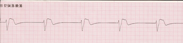
Idioventricular Rhythm. The rate is 33. The rhythm is regular. There are no P waves before the QRS complexes. The QRS complex is wide, greater than .12 sec. A slow rhythm with wide QRS complexes and absent P waves is characteristic of an idioventricular rhythm. This might be a rhythm that you would see in a patient with PEA. Recall that with PEA there is a rhythm on the monitor but the patient will be pulseless. What is the immediate treatment for a patient that is pulseless and apenic? How do you confirm true pulselessnes in a patient with PEA? What two drugs are indicated in the treatment of PEA? What are some reversible causes?
2.
5.
3rd Degree Heart Block. The rate is 34. The rhythm is regular. The P waves are not associcated with the QRS complexes. The QRS complexes are wide, > .12 sec. A slow rhythm with complete AV dissociation is characteristic of a 3rd degree heart block or complete heart block. You might see this rhythm with anterior infarcts because the LAD perfuses the intraventricular septum below the AV junction. Two catecholamines that can be administered to this patient: Epinephrine and dopamine

2.

3.

4.

5.

6.

Answers
1.
|
| Idioventricular Rhythm |
Idioventricular Rhythm. The rate is 33. The rhythm is regular. There are no P waves before the QRS complexes. The QRS complex is wide, greater than .12 sec. A slow rhythm with wide QRS complexes and absent P waves is characteristic of an idioventricular rhythm. This might be a rhythm that you would see in a patient with PEA. Recall that with PEA there is a rhythm on the monitor but the patient will be pulseless. What is the immediate treatment for a patient that is pulseless and apenic? How do you confirm true pulselessnes in a patient with PEA? What two drugs are indicated in the treatment of PEA? What are some reversible causes?
2.
|
| Bradycardia with Sinus Arrhythmia |
Bradycardia with Sinus Arrhythmia. The heart rate is 39. The rhythm is irregular. There are upright P waves before each QRS complexes. The PR interval is .16 sec. Because of the irregular rhythm this makes it a bradycardia with sinus arrhythmia. If the patient experienced this rhythm during insertion of an NG tube and was becoming more symptomatic, what drug could you administer the patient?
3.
|
| 2nd Degree Heart Block Type II |
2nd Degree Heart Block Type II. The heart rate is 40. The rhythm is irregular. There are upright P waves before each QRS complexes and there are some extra, nonconducted P waves present. The PR interval is .16 sec. Because of the extra P waves and the conducted P waves that have a consistent PR interval this rhythm is a 2nd degree heart block type II or Mobitz II heart block. This is a block that occurs below the level of the AV junction. You might see this rhythm with anterior infarcts because the LAD perfuses the intraventricular septum.
4.
|
| Sinus Bradycardia |
Sinus Bradycardia. The rate is 42. The rhythm is regular. There are upright P waves before each QRS complexes with a PR interval is .16 sec. The QRS complex is narrow, less than .12 sec. Based on this information, the rhythm is correctly identified as sinus bradycardia.
5.
|
| Junctional Rhythm |
Junctional Rhythm. The rate is 39. The rhythm is regular. There are inverted P waves before the QRS complexes. The PR interval is shortened. The QRS complex is narrow, < .12 sec. A slow rhythm with narrow complexes and with absent or inverted P waves is characteristic of a junctional rhythm. The patient's BP is 86/39. He is alert but mildly confused and anxious. He is holding his chest and is complaining of chest pain and is short of breath. His pulse oximetery is 92% on 2L/min via nasal cannula. His capillary refill is delayed, < 3 sec. His skin is cold and clammy. Is this patient stable or unstable? What is the recommended treatment for this patient?
6.
|
| 3rd Degree Heart Block |
Reviewed on 2/28/16


Comments
Post a Comment