EKG Rhythm Strips 14: Junctional Rhythms
1.

.JPG)
3.


5.


10.

Answers
1.
Junctional rhythm. The rhythm is regular. There are P waves but they are inverted and the QRS complexes are narrow (< .12 sec). What is the rate? This is the factor for determining which junctional rhythm you are looking at. Junctional rhythms arise from around the AV junction. The inherent rate of AV junctional area is 40-60 and the rhythm is called a junctional rhythm. When the rate is between 60-100 it is called an accelerated junctional rhythm and it is called junctional tachycardia when the rate exceeds 100 beats per minute. The characteristics of a junctional rhythm is that the rhythm is regular. The P wave may be absent or be present but inverted and precede or follow the QRS complex. The QRS complexes are usually narrow.
2.
Junctional Tachycardia. The rhythm is regular. What is the rate? Recall that you can either count the number of QRS complexes in a 6 second strip and mulitiply that number by 10 in order to determine the rate. Each small square is 0.4seconds. Each larger square is 0.2 seconds. So 5 large squares equals one second. The small hash marks at the bottom of the strip represents 3 seconds. Now that you know how to determine the rate, what is the rate and what is the rhythm?
3.
Junctional Rhythm. Actually this rhythm strip is a little slower than a normal junctional rhythm. But anything is possible when dealing with a sick heart. But it shows some good characteristics of a junctional rhythm. The inherent rate of the junctional tissue is______? The junctional tissue is inferior to the SA node and the atrium in the conduction pathway. So in order to activate the atrium, the impulse arising from the AV junction has to travel northward (antegrade). On the electocardiogram this will appear as a negative (inverted) P wave. If the atrium are depolarized before the ventricles then the inverted P wave will appear before the QRS complex. If the atrium and ventricles are depolarized at the same time, then the P wave will be absent. If the atrium are depolarized after the ventricles then the inverted P wave will appear after the QRS complex. In this rhythm strip the P wave follows the S wave (the small hump at the begining of the T wave). The P wave appears upright but this is not the case. As the T wave begins to rise and become more positive, you will see that it is interupted by a negative deflection, this is the negative P wave.
4.
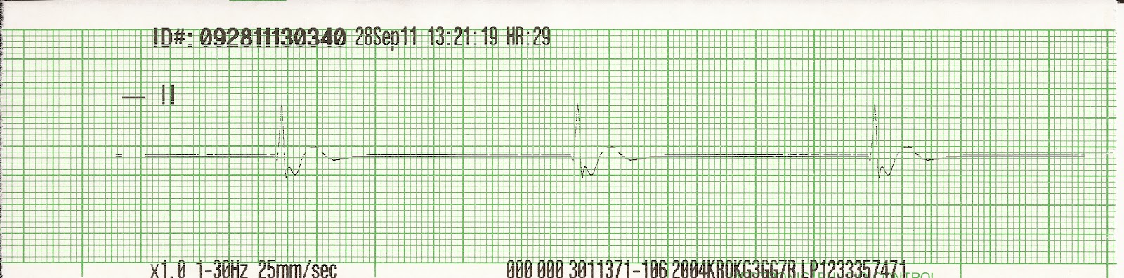
Junctional Rhythm. What is the rate? Is the atrium or ventricle depolarized first? This is a slow rhythm and the patient may be symptomatic or unstable or even pulseless. If the patient is pulseless, proceed using the PEA alorithm. If the patient is unstable: think immediate transcutaneous pacing. While setting up for pacing the MD may try Atropine 0.5mg or a catecholamine infusion with Dopamine or Epinephrine. What is the dosage range for Dopamine? Dopamine is a positive inotrophic agent will dilate the renal and messentary arteries at lower dose and cause increase myocardial contractility and vasoconstriction at higher doses. Thus, the MD might have you start at a dose of 5mcg/kg/min or higher in order to increase cardiac ouput and heart rate.
5.
Junctional Tachycardia. The rate is 125 so this puts it in the range of being a junctional tachycardia. The ventricles are depolarized first so the inverted P wave follows the QRS complex. A patient will not usually begin to be symptomatic until a heart rate gets over 150 (there are always exceptions). In general a junctional rhythm does not respond to cardioversion. Unlike SVT where the mechanism is a re-entry problem, a junctional rhythm arises from an escape mechanism. Whereas cardioversion can be used to interupt and break the re-entry cycle, it generally has no effect on rhythms that ectopic in nature. What is another fast ectopic rhythm? Hint: The rate is over 100 and it has a least 3 P waves of varying morphology? Hmmmm?
6.
Accelerated Junctional Rhythm. The rhythm is regular. There are no P waves. The QRS complex is narrow. Based upon the rate (what is the rate?) it is called an accelerated junctional rhythm. Where are the P waves? (See answer for rhythm 3 above) Notice that the QRS complex is narrow. This occurs because the ventricles are depolarized along the normal conduction pathway. If the QRS complexes in this rhythm strip were wide (>.12 sec) then you would probably say that the rhythm was ventricular in orgin. In which case it would be called an accelerated ______ rhythm? Hint: it is characterized by a wide QRS complex with a rate between 40 and 100).
7.
Accelerated Junctional Rhythm. Based upon the rate of 69, this rhythm is properly identified as an accelerated junctional rhythm. There are P waves present and they are inverted before the QRS complex. They almost resemble delta waves. In what rhythm are delta waves associated with?
8.
Accelerated Junctional Rhythm. The rhythm is regular. The rate is 88. The P waves are inverted and the QRS complex is narrow. The inverted P waves suggests a rhythm that is junctional in nature.
9.
Junctional Rhythm. The rhythm is regular. The P waves are inverted and follow the QRS complex. Based upon the slow rate, the rhythm is properly identified as a junctional rhythm. If the patient were hypotensive, cold and clammy, confused, short of breath, and had chest pain, how would you treat the patient?
10.
Junctional Tachycardia. The rhythm is regular. The P waves are inverted and follow the QRS complex. The QRS complex is narrow. Because the rate is > 100, this rhythm is readily identified as junctional tachycardia. As the rate approaches 150 the QRS complexes get closer and closer together and the P waves may not be evident at all. If the patient were symptomatic only, you might try some vagal manuvers in order to treat or identify the rhythm. Recall that vagal maneuvers are both theraputic and diagnostic. What are 5 vagal maneuvers you could try? What are the restrictions for using carotid sinus massage?
Reviewed 2/28/16

2.
.JPG)

4.

5.

6.


7.

8.

9.
10.

Answers
1.
|
| Junctional rhythm |
Junctional rhythm. The rhythm is regular. There are P waves but they are inverted and the QRS complexes are narrow (< .12 sec). What is the rate? This is the factor for determining which junctional rhythm you are looking at. Junctional rhythms arise from around the AV junction. The inherent rate of AV junctional area is 40-60 and the rhythm is called a junctional rhythm. When the rate is between 60-100 it is called an accelerated junctional rhythm and it is called junctional tachycardia when the rate exceeds 100 beats per minute. The characteristics of a junctional rhythm is that the rhythm is regular. The P wave may be absent or be present but inverted and precede or follow the QRS complex. The QRS complexes are usually narrow.
2.
 |
| Junctional Tachycardia |
Junctional Tachycardia. The rhythm is regular. What is the rate? Recall that you can either count the number of QRS complexes in a 6 second strip and mulitiply that number by 10 in order to determine the rate. Each small square is 0.4seconds. Each larger square is 0.2 seconds. So 5 large squares equals one second. The small hash marks at the bottom of the strip represents 3 seconds. Now that you know how to determine the rate, what is the rate and what is the rhythm?
3.
|
| Junctional Rhythm |
Junctional Rhythm. Actually this rhythm strip is a little slower than a normal junctional rhythm. But anything is possible when dealing with a sick heart. But it shows some good characteristics of a junctional rhythm. The inherent rate of the junctional tissue is______? The junctional tissue is inferior to the SA node and the atrium in the conduction pathway. So in order to activate the atrium, the impulse arising from the AV junction has to travel northward (antegrade). On the electocardiogram this will appear as a negative (inverted) P wave. If the atrium are depolarized before the ventricles then the inverted P wave will appear before the QRS complex. If the atrium and ventricles are depolarized at the same time, then the P wave will be absent. If the atrium are depolarized after the ventricles then the inverted P wave will appear after the QRS complex. In this rhythm strip the P wave follows the S wave (the small hump at the begining of the T wave). The P wave appears upright but this is not the case. As the T wave begins to rise and become more positive, you will see that it is interupted by a negative deflection, this is the negative P wave.
4.
|
| Junctional Rhythm |
Junctional Rhythm. What is the rate? Is the atrium or ventricle depolarized first? This is a slow rhythm and the patient may be symptomatic or unstable or even pulseless. If the patient is pulseless, proceed using the PEA alorithm. If the patient is unstable: think immediate transcutaneous pacing. While setting up for pacing the MD may try Atropine 0.5mg or a catecholamine infusion with Dopamine or Epinephrine. What is the dosage range for Dopamine? Dopamine is a positive inotrophic agent will dilate the renal and messentary arteries at lower dose and cause increase myocardial contractility and vasoconstriction at higher doses. Thus, the MD might have you start at a dose of 5mcg/kg/min or higher in order to increase cardiac ouput and heart rate.
5.
|
| Junctional Tachycardia |
Junctional Tachycardia. The rate is 125 so this puts it in the range of being a junctional tachycardia. The ventricles are depolarized first so the inverted P wave follows the QRS complex. A patient will not usually begin to be symptomatic until a heart rate gets over 150 (there are always exceptions). In general a junctional rhythm does not respond to cardioversion. Unlike SVT where the mechanism is a re-entry problem, a junctional rhythm arises from an escape mechanism. Whereas cardioversion can be used to interupt and break the re-entry cycle, it generally has no effect on rhythms that ectopic in nature. What is another fast ectopic rhythm? Hint: The rate is over 100 and it has a least 3 P waves of varying morphology? Hmmmm?
6.
|
| Accelerated Junctional Rhythm |
Accelerated Junctional Rhythm. The rhythm is regular. There are no P waves. The QRS complex is narrow. Based upon the rate (what is the rate?) it is called an accelerated junctional rhythm. Where are the P waves? (See answer for rhythm 3 above) Notice that the QRS complex is narrow. This occurs because the ventricles are depolarized along the normal conduction pathway. If the QRS complexes in this rhythm strip were wide (>.12 sec) then you would probably say that the rhythm was ventricular in orgin. In which case it would be called an accelerated ______ rhythm? Hint: it is characterized by a wide QRS complex with a rate between 40 and 100).
7.
|
| Accelerated Junctional Rhythm |
Accelerated Junctional Rhythm. Based upon the rate of 69, this rhythm is properly identified as an accelerated junctional rhythm. There are P waves present and they are inverted before the QRS complex. They almost resemble delta waves. In what rhythm are delta waves associated with?
8.
|
| Accelerated Junctional Rhythm |
Accelerated Junctional Rhythm. The rhythm is regular. The rate is 88. The P waves are inverted and the QRS complex is narrow. The inverted P waves suggests a rhythm that is junctional in nature.
9.
|
| Junctional Rhythm |
Junctional Rhythm. The rhythm is regular. The P waves are inverted and follow the QRS complex. Based upon the slow rate, the rhythm is properly identified as a junctional rhythm. If the patient were hypotensive, cold and clammy, confused, short of breath, and had chest pain, how would you treat the patient?
10.
|
| Junctional Tachycardia |
Junctional Tachycardia. The rhythm is regular. The P waves are inverted and follow the QRS complex. The QRS complex is narrow. Because the rate is > 100, this rhythm is readily identified as junctional tachycardia. As the rate approaches 150 the QRS complexes get closer and closer together and the P waves may not be evident at all. If the patient were symptomatic only, you might try some vagal manuvers in order to treat or identify the rhythm. Recall that vagal maneuvers are both theraputic and diagnostic. What are 5 vagal maneuvers you could try? What are the restrictions for using carotid sinus massage?
Reviewed 2/28/16


Comments
Post a Comment