Practice EKG Rhythm Strips 219
Identify the following rhythms.
1.
2.
3.
4.
5.
Answers
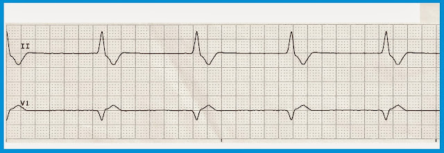
1.
The rhythm is regular with a rate of 48/min. No P waves are seen. The QRS is wide, suggesting a ventricular origin for the rhythm. There are no ectopic beats. PR: ---, QRS: .12 sec, QT: .38 sec. Interpretation: Accelerated idioventricular rhythm
2.
The rhythm is irregular. The rate is 90/min. There are upright P waves associated with a QRS complex. The are two unifocal PVCs present. PR: .16 sec, QRS: .08 sec, QT: .38 sec. Interpretation: Normal sinus rhythm with unifocal PVCs
3.
The rhythm is regular with a rate of 57/min. P waves are present but there are not corresponding QRS complexes, there is no ventricular activity. Interpretation: P wave asystole or ventricular standstill.
4.
The rhythm is regular with a rate of 62/min. No P waves are seen, just some fibrillation between the QRS complexes. A ventricular pacer spike precedes each QRS complex. No ectopic beats are noted. PR: ---, QRS: .16 sec, QT: .44 sec. Interpretation: Ventricular paced rhythm
5.
The rhythm is regular. The rate is 136/min. No P waves are seen. The QRS complex is wide which points toward a ventricular origin for this rhythm. No ectopic beats are noted. PR: ---, QRS: .16 sec, QT: .36 sec. Interpretation: Ventricular tachycardia.
1.
2.
3.
4.
5.
Answers
1.
 |
| Accelerated idioventricular rhythm |
The rhythm is regular with a rate of 48/min. No P waves are seen. The QRS is wide, suggesting a ventricular origin for the rhythm. There are no ectopic beats. PR: ---, QRS: .12 sec, QT: .38 sec. Interpretation: Accelerated idioventricular rhythm
2.
 |
| Normal sinus rhythm with unifocal PVCs |
The rhythm is irregular. The rate is 90/min. There are upright P waves associated with a QRS complex. The are two unifocal PVCs present. PR: .16 sec, QRS: .08 sec, QT: .38 sec. Interpretation: Normal sinus rhythm with unifocal PVCs
3.
 |
| P wave asystole |
The rhythm is regular with a rate of 57/min. P waves are present but there are not corresponding QRS complexes, there is no ventricular activity. Interpretation: P wave asystole or ventricular standstill.
4.
 |
| Ventricular paced rhythm with underlying atrial fibrillation |
The rhythm is regular with a rate of 62/min. No P waves are seen, just some fibrillation between the QRS complexes. A ventricular pacer spike precedes each QRS complex. No ectopic beats are noted. PR: ---, QRS: .16 sec, QT: .44 sec. Interpretation: Ventricular paced rhythm
5.
 |
| Ventricular tachycardia |
The rhythm is regular. The rate is 136/min. No P waves are seen. The QRS complex is wide which points toward a ventricular origin for this rhythm. No ectopic beats are noted. PR: ---, QRS: .16 sec, QT: .36 sec. Interpretation: Ventricular tachycardia.


Comments
Post a Comment