Posts

Showing posts with the label Escape beats

Atrial Fibrillation with Ventricular Escape Beats

Image
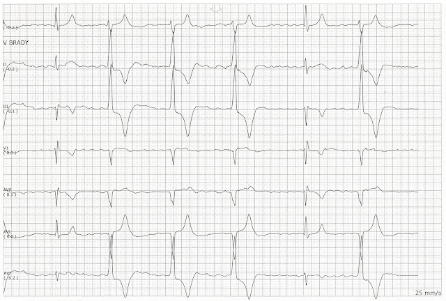
Atrial Fibrillation with Ventricular Escape Beats.  These are two consecutive pages on the same patient.  She was in an atrial fibrillation with slow ventricular response.   She began having some runs of this ventricular escape rhythm.   

Sinus bradycardia changing to a ventricular escape rhythm

Image
Sinus bradycardia changing to a ventricular escape rhythm  

Junctional escape beats

Image
A dropped PAC is seen after the 2nd complex.  This is followed by three junctional escape beats.  A PAC occurs (6th complex) and another junctional escape beat is seen (7th complex)  

Dropped PACs and escape beats

Image
Dropped PACs.   There is an underlying first degree block  A dropped PAC occurs after the 5th complex.  The dropped P wave can be seen better in the AVF lead.  This is followed by an atrial escape beat (6th complex) and a junctional escape beat (7th complex).  A PVC (8th complex) is next. The 9th complex is a junctional escape beat.  A dropped PAC follows.  The 10th complex is another junctional escape complex.   

First degree block with some ventricular escape beats

Image
First degree block with some ventricular escape beats.   In lead II a PVC (2nd complex) is seen.  The PR interval of the 3rd complex is prolonged.  The 4th complex is sinus with a first degree block.  The 5th complex is a PVC.  A dropped beat follows the PVC.  You can see it better in lead I.  This is followed by 4 ventricular escape beats.  In lead I it looks like some dissociated P waves are present with the escape beats.   

Atrial fibrillation with some ventricular escape beats

Image
Atrial fibrillation with a slow ventricular response and escape beats.   The QRS complexes are wide in the underlying rhythm.  Complexes 1 - 3 show the patient's underlying heart rhythm.  Complexes 4 and 5 are ventricular escape beats that arrive later in the cardiac cycle.   

Sinus bradycardia with ventricular escape beats

Image
In these two pages the patient is having sinus bradycardia with ventricular escape beats.  The underlying rhythm is sinus bradycardia.  The 5th complex is a PVC.  It is followed by two ventricular escape beats.  Sinus bradycardia resumes after the escape beats.   

Sinus Rhythm with Ventricular Escape Beats

Image
The rhythm is irregular.  An upright P wave is paired with a QRS complex.  The PR interval is prolonged. We have multifocal PVCs present. A pause follows the PVCs.  The 2nd PVC is followed by some ventricular escape beats.  

Multiple and Consecutive PACs with Junctional Escape Beats

Image
The are multiple and consecutive PACs present.  It even looks like there is a dropped PAC that follows the 1st,6th, 12th, and 13th complexes.  The 1st, 7th, 13th, and 14th complexes look like junctional escape beats. 

Sinus Bradycardia with Ventricular Escape Beats

Image
On this page the patient is in a sinus bradycardia.  There is a PVC after the third complex and this is followed by two ventricular escape beats.

EKG Rhythm Strip

Image
Identify the following rhythm. (Click on the image to enlarge) Atrial fibrillation with ventricular escape beats.  The underlying rhythm is atrial fibrillation.  Beats  4, 5, and 6 occur late in the cardiac cycle. The morphology of the QRS complex is much wider than the QRS complexes of beats 1, 2, 3, and 7. The R - R interval between beats 3 and 4 is about 37 small boxes, this corresponds to a rate of about 40 beats/min.  Since the inherent rate of the ventricles is 20 - 40 bpm, complex 4 falls within the upper limits of an escape beat of ventricular origin. Complexes 5 and 6 occur at the same rate.  

Various Escape Beats

Image
Various Escape Beats 1. Sinus Bradycardia with Ventricular Escape Beats 2. First degree block with a dropped complex followed by a ventricular escape beat 3. Atrial fibrillation with ventricular escape beats 4. Sinus bradycardia with some ventricular escape beats 5. Sinus bradycardia with ventricular escape beats 6. Sinus bradycardia with bigeminal junctional escape beats 7. Sinus arrhythmia with a dropped PAC and a ventricular escape beat 8. Atrial fibrillation with ventricular escape beats 9. Sinus rhythm with a PVC followed by junctional escape beats 10. Sinus rhythm with a junctional escape beat 11. Sinus rhythm with a PVC followed by ventricular escape beats

Practice EKG Rhythm Strips 215

Image
Identify the following rhythms. 1. 2. 3. 4. 5. Answers 1. 2nd degree AV block type I The rhythm is irregular with a rate of 60/min.   There are upright P waves present.   There is progressive elongation of the PR interval over successive beats.   A non-conducted P wave follows the 4th complex.   No ectopic beats are present.   The QRS complex is narrow.   PR:  .28/.32/.36 sec.  QRS:  .08 sec,  QT:  .44 sec.   Interpretation:  2nd degree AV block type I 2. Atrial paced rhythm  The rhythm is regular with a rate of 75/min.   There are small upright P waves that follow the atrial pacer spike.  No ectopic beats are seen.  PR:  .28 sec.  QRS:  .08 sec,  QT:  .34 sec.   Interpretation:  Atrial paced rhythm 3. NSR with a pause followed by a junctional escape beat  The rhythm is irregular.   The rate is 70/min.   The P waves are uniform, positive, and have a corresponding QRS complex.   A pause follows the 3rd complex.  

Practice EKG Rhythm Strips 201

Image
Identify the following rhythms. 1. 2. 3. 4. 5. Answers 1. 1st degree block with a PVC The rhythm is irregular due to the PVC.   The rate is 60/min.    The P waves are upright and precede the QRS complexes.   The PR interval is prolonged.   There is a single PVC present.  PR:  .36 sec,  QRS:  .08 sec,  QT:  .40 sec. 2. Agonal rhythm The rhythm may be regular or irregular.   The rate is 20/min.    No P waves are present.   The QRS complexes are wide suggesting a ventricular origin to the rhythm.   PR:  ---,  QRS:  .20 sec,  QT:  .52 sec. 3. Atrial fibrillation  The rhythm is irregular with a heart rate of 100/min.   The P waves are absent.   There are flutter waves seen between the QRS complex,  seen more clearly in the V1 lead.  No ectopic beats are noted.  PR:  ---,  QRS:  .08 sec,  QT:  .32 sec. 4. Complete heart block  The rhythm is regular with a ventricular rate of 34 and an atrial rate of 100/min.   The P w

Practice EKG Rhythm Strips 160

Image
Identify the following rhythms. 1. 2. 3. 4. 5. Answers 1. Normal sinus rhythm The rhythm is regular with a calculated rate of 78/min.   There are uniform, upright P waves before each QRS complex.  No ectopy is noted.   PR:  .12,  QRS,  .08 sec,  QT:  .28 sec. 2. Normal sinus rhythm with PACs The rhythm is irregular due to some PACs.   The rate is 90/min.   There are upright P waves before each QRS complex.   The P waves are not uniform in shape.   There are some atrial ectopic beats present:  complexes 3, 5, and 11.  The R-R interval on these beats is shorter than the surrounding sinus beats.  In addition the P wave morphology and the PR intervals also differ.  PR:  .12 sec,  QRS:  .08 sec,  QT:  .32 sec. 3. Normal sinus rhythm with PVCs The rhythm is irregular due to the PVCs.   The rate is 70/min.   The P waves are upright and uniform on the sinus beats.   There are two PVCs of slightly different