Posts

Showing posts with the label Atrial Fibrillation

Atrial fibrillation with periods of arrest

Image
Atrial fibrillation with periods of arrest  

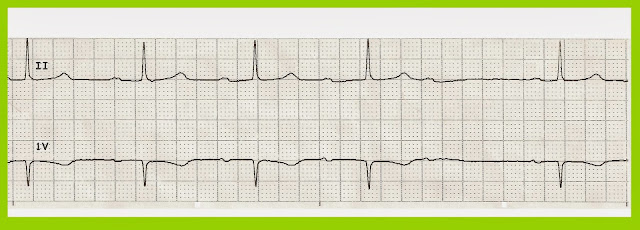
Atrial fibrillation with slow ventricular response

Image
Atrial fibrillation with slow ventricular response.  In the V1 lead it looks more like an atrial flutter.  The conduction through the AV node is variable and slow.   

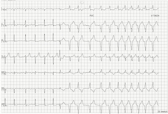
Atrial fibrillation changing to ventricular tachycardia

Image
These two pages show are a continuous rhythm of atrial fibrillation changing to ventricular tachycardia.     

Atrial fibrillation with a slow ventricular response and arrest

Image
Atrial fibrillation with a slow ventricular response and a long period of arrest  

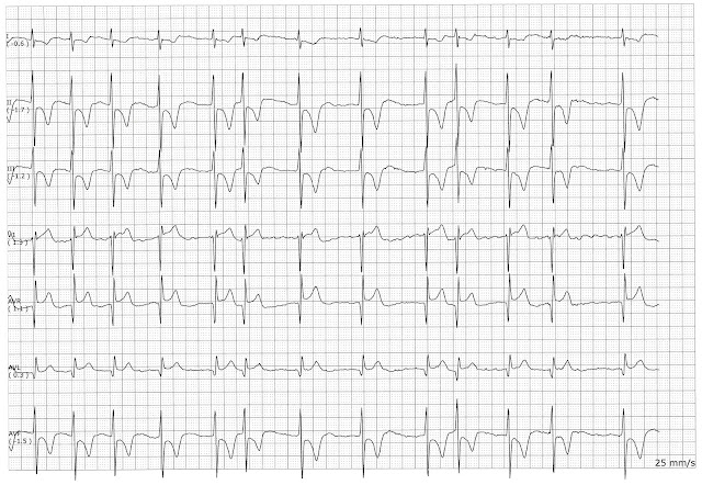
Atrial Fibrillaion

Image
The underlying rhythm is atrial fibrillation.  there is also T wave inversion in the inferior leads II, III, and aVF.   

Atrial fibrillation with a Run of Ventricular Tachycardia

Image
This page shows a patient in atrial fibrillation who had a nonsustained run of VT.  The patient converted out of the VT without any intervention.

Atrial Fibrillation with Slow Ventricular Response

Image
On this page the page has atrial fibrillation with slow ventricular response.  We are more accustomed to seeing atrial fibrillation with a rapid ventricular response.  However, when the patient takes too much medication or has an electrolyte problem we can see a slower atrial fibrillation.

Ventricular Bigeminy

Image
On this page, the patient is in atrial fibrillation and is having some bigeminal PVCs

Various Atrial Fibrillations

Image
Various Atrial Fibrillations 1. Atrial Fibrillation with RVR 2. Atrial Fibrillation with Multifocal PVCs 3. Atrial Fibrillation with Multifocal PVCs 4. Atrial Fibrillation 5. Atrial Fibrillation 6. Atrial Fibrillation 7. Atrial fibrillation with slow ventricular response 8. Atrial fibrillation with rapid ventricular response 9. Atrial fibrillation with controlled rate 10. Atrial fibrillation with rapid ventricular response 11. Atrial fibrillation with rapid ventricular response 12. Atrial fibrillation with rapid ventricular response 13. Atrial fibrillation with 4 beat run of VT 14. Atrial fibrillation with rapid ventricular response 15.

Practice EKG Rhythm Strips 220

Image
Identify the following rhythms. 1. 2. 3. 4. 5. Answers 1. Sinus arrhythmia with 1st degree block The rhythm is irregular with a rate of 70/min.   The P waves are upright, uniform, and precede the QRS complexes.  The P waves are split which suggests biatrial enlargement.  The PR interval is prolonged.  The QRS is narrow.  No ectopic beats are noted.   PR:  .24 sec,  QRS:  .08 sec,  QT:  .44 sec.   Interpretation:   Sinus arrhythmia with 1st degree block 2.  Idioventricular rhythm The rate is 20/min.  There are no P waves.   The QRS complex is wide which is indicative of a ventricular escape rhythm.  PR:  ---,  QRS:  .18 sec,  QT:  .50 sec.   Interpretation:  Idioventricular rhythm 3.  Atrial fibrillation with multifocal PVCs The rhythm is irregular with a heart rate o...

Practice EKG Rhythm Strips 210

Image
Identify the following rhythms. 1. 2. 3. 4. 5. Answers 1. 2nd degree AV block type I The rhythm is irregular with a rate of 50/min.   The P waves are upright and uniform.   There is gradual elongation of the PR interval.    There is a nonconducted P wave after the 4th complex.  No ectopic beats are seen.   The QRS complex is narrow.   PR:  .28/.32/.36 sec.  QRS:  .08 sec, QT:  .48 sec.  Interpretation:  2nd degree AV block type I 2. Atrial fibrillation  The rhythm is irregular with a rate of 80/min.   No P waves are seen.  There is an aberrantly conducted complex that arrives later in the cycle after the 4th complex.  There is 3 mm ST elevation in the V1 lead.  PR:  ---,  QRS:  .12 sec,  QT:  .44 sec.   Interpretation:  Atrial fibrillation 3. Atrial paced The rhythm is regular ...

Practice EKG Rhythm Strips 205

Image
Identify the following rhythms. 1. 2. 3. 4. 5. Answers 1. 1st degree AV block with a PVC The rhythm is irregular with a PVC.   The heart rate is 60/min.    The P waves are upright and are associated with a QRS complex.   The PR interval is prolonged.    A single PVC is present.   PR:  .44 sec.  QRS:  .16 sec,  QT:  .50 sec. 2. Agonal rhythm The rhythm is irregular.    The rate is 20/min.   No P waves are seen.   The QRS complex is wide suggesting a rhythm of ventricular origin.   PR:  ---,  QRS:  .20 sec,  QT:  .56 sec.   3. Atrial fibrillation with slow ventricular response The rhythm is irregular with a rate of 40/min.    No P waves are seen.   Some fibrillation is present...