Posts

Showing posts with the label Atrial paced

Practice EKG Rhythm Strips 217

Image
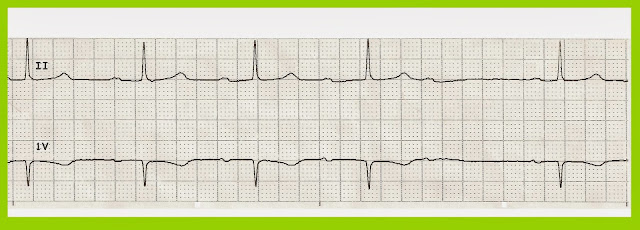
Identify the following rhythms. 1. 2. 3. 4. 5. Answers 1. 2nd degree heart block type II The rhythm is irregular.   The rate is 30/min.  There are upright, uniform P waves present but not all of them are associated with a QRS complex.  A non-conducted P wave follows each QRS complex.  The PR interval is constant on the conducted P waves.  There are two PVCs present, the 1st complex and the 4th complex.   PR:  .36 sec,  QRS:  .12 sec,  QT:  .48 sec.   Interpretation: 2nd degree heart block type II 2. Atrial flutter The rhythm is irregular with a rate of 90.   Flutter waves are present.  No ectopic beats are seen.  PR:---,  QRS:  .12 sec,  QT:.44 sec.   Interpretation:  Atrial flutter. 3. Atrial paced The rhythm is regular with a rate of 75/min.   A small P wave follows the atrial pacer spike.   No ectopic beats are seen.  PR:  .28 sec,  QRS:  .12 sec,  QT:
Practice EKG Rhythm Strips 210

Image
Identify the following rhythms. 1. 2. 3. 4. 5. Answers 1. 2nd degree AV block type I The rhythm is irregular with a rate of 50/min.   The P waves are upright and uniform.   There is gradual elongation of the PR interval.    There is a nonconducted P wave after the 4th complex.  No ectopic beats are seen.   The QRS complex is narrow.   PR:  .28/.32/.36 sec.  QRS:  .08 sec, QT:  .48 sec.  Interpretation:  2nd degree AV block type I 2. Atrial fibrillation  The rhythm is irregular with a rate of 80/min.   No P waves are seen.  There is an aberrantly conducted complex that arrives later in the cycle after the 4th complex.  There is 3 mm ST elevation in the V1 lead.  PR:  ---,  QRS:  .12 sec,  QT:  .44 sec.   Interpretation:  Atrial fibrillation 3. Atrial paced The rhythm is regular with a rate of 65/min.  The are upright P waves that are preceded by atrial pacer spikes.   The PR interval is prolonged.  T

Practice EKG Rhythm Strips 203

Image
Identify the following rhythms. 1. 2. 3. 4. 5. Answers 1. Accelerated junctional rhythm The rhythm is regular with a heart rate of 71/min.   The P waves are absent.   No ectopic beats are noted.   The QRS complex is narrow.   PR:  ---,  QRS:  .08 sec,  QT:  .40 sec. 2. Multifocal atrial tachycardia The rhythm is irregular with ventricular rate of 150/min.    P waves are upright but are not uniform in appearance.   There is also some variability in the PR interval.   No ectopic beats are noted.  PR:  .16 sec,  QRS:  .08 sec,  QT:  .32 sec. 3. Atrial flutter The rhythm is regular with a rate of 71/min.   There flutter waves seen between the QRS complexes.  No ectopic beats are noted.  PR:  ---,  QRS:  .08 sec,  QT:  undetermined. 4. Atrial paced The rhythm is regular with a heart rate of 68/min.   The P waves are preceded by an atrial pacing stimulus.   No ectopic beats are seen.   PR:  .20

Practice EKG Rhythm Strips 182

Image
Identify the following rhythms. 1. 2. 3. 4. 5. Answers 1. Atrial fibrillation with rapid ventricular response The rhythm is very irregular with a rate of 160/min.  No P waves are seen.  No ectopic beats are noted. PR:  ---,  QRS:  .10 sec,  QT:  .28 sec. 2. Atrial paced The rhythm is regular with a heart rate of 68/min.   The P waves are preceded by an atrial pacing stimulus.   No ectopic beats are noted.   PR:  .16 sec,  QRS:  .08 sec,  QT:  .36 sec. 3. Dual paced The rhythm is regular.   The heart rate is 68/min.   The P waves and the QRS complexes are preceded by a pacer spike.   PR:  .12 sec,  QRS:  .16 sec,  QT:  .44 sec. 4. Normal sinus rhythm with bigeminal PVCs The rhythm is irregular due to the multiple PVCs.   The ventricular rate is 100/min.   The P waves are uniform, positive, and are associated with a QRS complex.   There are unifocal PVCs every other

Practice EKG Rhythm Strips 162

Image
Identify the following rhythms. 1. 2. 3. 4. 5. Answers 1. Atrial paced This paced rhythm is regular with a rate of 60/min.  There are upright P waves before each QRS complexes.   Each P wave is preceded by a pacer spike.   No ectopic beats are noted.  PR:  .20 sec,  QRS:  .08 sec, QT .48 seconds. 2. Normal sinus rhythm with ST elevation in lead II The rhythm is regular and the rate is 88/min.  The P waves are associated with the QRS complexes and are uniform and upright.   No ectopic beats are seen. In lead II there is 2mm ST elevation with some inversion of the T wave.  PR:  .16 sec,  QRS:  .08 sec,  QT:  .40 sec. 3. Normal sinus rhythm with a PAC This rhythm is irregular due to the PAC.   The rate is 80/min.   The P waves are upright and precede the QRS complex.  The 3rd complex is a PAC.  Notice the change in the height of the preceding T wave.   The change in height is the