Posts

Showing posts with the label Dual paced

Pacemaker Problems

Image
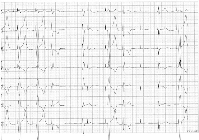
This strips shows an AV paced rhythm.   After the 4th complex there is failure to capture.  The fifth complex show failure to sense.  The AV interval on the sixth complex is shorter than the previous paced beats.  Between the 7th and 8th complex there is failure to capture.  The 8th complex is a fusion complex.

Practice EKG Rhythm Strips 211

Image
Identify the following rhythms. 1. 2. 3. 4. 5. Answers 1.  Accelerated junctional rhythm.   The rhythm is regular with a rate of 94/min.   No P waves are seen.   No ectopic beats are seen. The QRS complex is wide.   PR:  ---,  QRS:  .12 sec,  QT:  .40 sec.  Interpretation:  Accelerated idioventricular rhythm.   The interpretation of accelerated idioventricular rhythm is correct based upon the characteristic of the rhythm.  However, this patient actually had an underlying wide QRS complex on previous strips so the actual interpretation for this strip would be an accelerated junctional rhythm. 2. Asystole with a functional pacemaker  There are no P waves are QRS complexes.   There are pacer spikes present but they are not capturing.   Interpretation:  Asystole with a functional pacemaker 3. Dual paced with biventricula...

Practice EKG Rhythm Strips 200

Image
Identify the following rhythms. 1. 2. 3. 4. 5. Answers 1. Sinus bradycardia with a PJC The rhythm is irregular with a heart rate of 50/min.   The P waves are upright, except for the 2nd complex.   The P wave of the PJC is negative.   PR:  .16 sec,  QRS:  .08 sec, QT:  .40 sec. 2. Ventricular paced with unifocal PVCs The rhythm is irregular due to the PVCs.   The ventricular rate is 60/min.   No P waves are seen. There are two unifocal PVCs present, the 1st and 4th complexes.   There are ventricular pacer spikes before the remaining QRS complexes.  PR:  ---,  QRS:  .16 sec,  QT:  .44 sec. 3. Junctional tachycardia The rhythm is regular with a rate of 125/min.   The P waves are absent.  No ectopic beats are noted.  The QRS complex is narrow.   PR:  ---,  QRS:  .08 sec,  QT...
Practice EKG Rhythm Strips 132

Image
Identify the following rhythms. 1. 2. 3. 4. 5. Answers 1. Atrial flutter with variable block The rhythm is irregular.   The rate is 50/min.    No P waves are present, only flutter waves.   The first few complexes have a 3:1 ratio of flutter waves to QRS complexes.  Then it is followed by a 13 second arrest period that is dominated by the flutter waves.   No ectopy is present.  PR: --,  QRS:  .08 sec,  QT:  28 sec.  It is sometime difficult to measure the QT interval in atrial flutter because sometimes the flutter waves are buried within the T waves. 2. Demand pacing The rhythm is fairly regular.   The calculated rate is 65/min.   The first four complexes have a small upright sinus P waves.   The 6th and 7th complexes have an atrial paced P wave.  The 5th complex is a ventricular ectopic beat.  The last two complexes are dual paced.  P...

Practice EKG Rhythm Strips 127

Image
Identify the following rhythms. 1. 2. 3. 4. 5. Answers 1. Biventricular paced  I don't see a P wave after the 1st pacer spike and the pacer spikes occur just prior to the QRS complexes.  The rhythm is regular.   The rate is 71/min.    Very small upright P waves are noted before each QRS complex.  No ectopic beats are present.  A biventricular pacer spike preceeds each QRS complex. 2. Atrial fibrillation with rapid ventricular response The rhythm is irregular.   The rate is 140/min.   There are no P waves present,  fibrillatory waves only.   No ectopic beats are noted.   PR:  ---,  QRS:  .06 sec,  QT:  .38 sec. 3. Normal sinus rhythm The rhythm is regular.   The calculated rate is 93/min.   The P waves are upright and precede each QRS complex.   No ectopic beats are identified.   PR.  12 sec,  QRS: ...

Practice ECG Rhythm Strips 114

Image
Identify the following rhythms. 1. 2. 3. 4. 5. Answers 1. Accelerated junctional rhythm The rhythm is regular with a rate of 65/min.   There are negative P waves that precede each QRS complex.  No ectopic beats are noted.  PR:  .12 sec,  QRS:  .08 sec,  QT:  .40 sec. 2. Demand pacing with PACs  The rhythm is irregular with a rate of 80/min.  There are atrial and ventricular pacer spikes present on beats 1, 3, 5, and 7.   There are early beats every other beat which have only a ventricular pacer spike.  These beats appear to be PACs.   If you look at the 8th complex there is a P wave that precedes the QRS complex.  The 5th complex has a taller T wave than the other T waves which suggests that a P wave is buried within it. 3. Normal sinus rhythm with a dropped premature beat and escape beats The rhythm is irregular with a ...