Posts

Showing posts with the label Idioventricular rhythm

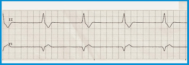
Idioventricular rhythm

Image
 Idioventricular rhythm  

Various Idioventricular Rhythms

Image
Various Idioventricular Rhythms 1. Idioventricular rhythm 2. Idioventricular rhythm 3. Idioventricular rhythm 4. Idioventricular rhythm 5. Idioventricular rhythm 6. Idioventricular rhythm 7. Idioventricular rhythm 8. Idioventricular rhythm 9. Idioventricular rhythm 10. Idioventricular rhythm 11. Idioventricular rhythm 12. Idioventricular rhythm 13. Idioventricular rhythm 14. Idioventricular rhythm 15. Accelerated idioventricular rhythm 16. Atrial flutter with run of AIVR 17.

Practice EKG Rhythm Strips 219

Image
Identify the following rhythms. 1. 2. 3. 4. 5. Answers 1. Accelerated idioventricular rhythm The rhythm is regular with a rate of 48/min.   No P waves are seen.  The QRS is wide, suggesting a ventricular origin for the rhythm.  There are no ectopic beats.  PR:  ---,  QRS:  .12 sec,  QT:  .38 sec.   Interpretation:  Accelerated idioventricular rhythm 2.  Normal sinus rhythm with unifocal PVCs The rhythm is irregular.  The rate is 90/min.  There are upright P waves associated with a QRS complex.   The are two unifocal PVCs present.   PR:  .16 sec,  QRS:  .08 sec,  QT:  .38 sec.   Interpretation:   Normal sinus rhythm with unifocal PVCs 3.  P wave asystole The rhythm is regular with a rate of 57/min.   P waves are present but there are not corresponding QRS complexes, the...

Practice EKG Rhythm Strips 213

Image
Identify the following rhythms. 1. 2. 3. 4. 5. Answers 1. Atrial fibrillation with a PVC The rhythm is irregular with a rate of 60/min.   No P waves are present.   Fibrillation and flutter waves are present between the QRS complexes.  The 5th complex is a PVC.  There is some ST depression in  lead II.  PR:  ---,  QRS:  .08 sec,  QT:  .36 sec.   Interpretation:  Atrial fibrillation with a PVC 2. The rhythm is regular.   The heart rate is 48/min.  No P waves are seen.  The QRS complex is wide suggesting a ventricular origin for this rhythm.  No ectopic beats are noted.  PR:  ---,  QRS:  .12 sec,  QT:  .36 sec.  Interpretation:  Accelerated idioventricular rhythm. 3. NSR with a PVC triplet The rhythm is irregular due to the PVCs.   The rate is 90/min.  There are uprig...

Practice EKG Rhythm Strips 198

Image
Identify the following rhythms. 1. 2. 3. 4. 5. Answers 1. Agonal rhythm The rhythm is irregular.  The rate is 20/min.  No atrial activity is present.   The QRS complex is wide, bizzare looking, and slow. 2. Biventricular paced The rhythm is regular with a heart rate of 94/min.   The P waves are uniform, upright, and precede the QRS complexes.   There are biventricular pacer spikes seen before each QRS complex.  No ectopic beats are noted.  PR:  .12 sec,  QRS: .12 sec,  QT:  .40 sec. 3. Atrial fibrillation with slow ventricular response The rhythm is irregular with a ventricular rate of 30/min.   The R-R interval between the 1st and 2nd complexes is 1.68 sec and the R-R interval between the 2nd and 3rd complex is 1.52 sec.  No P waves are present but there looks like some small flutter waves present in the V1 lead, t...

Practice EKG Rhythm Strips 192

Image
Identify the following rhythms. 1. 2. 3. 4. 5. Answers 1. Asystole with pacer spikes The pacemaker is still functioning in this asystolic rhythm. 2. Atrial flutter The heart rhythm is regular with a rate of 53/min.   There are flutter waves seen between the QRS complexes.   The atrial rate is around 200/min.  No ectopic beats are noted.  PR:  ---,  QRS:  .08 sec,  QT:  .44 sec. 3. Idioventricular rhythm The rhythm is regular with a rate of 35/min.  No P waves are seen.   The QRS is wide indicating a ventricular escape origin.   PR:  ---,  QRS:  .12 sec,  QT:  .36 sec. 4. Multifocal atrial tachycardia The rhythm is irregular with multiple PACs and a ventricular rate of 110/min.   The P waves are associated with a QRS complex but are not uniform.   There is also some variability in the PR interval.   PR:  . 12 s...