Posts

Showing posts with the label Sinus pause

Various Sinus Arrests and Sinus Pauses

Image
Various Sinus Arrests and Sinus Pauses 1. Sinus bradycardia with sinus arrest 2. Sinus bradycardia with a sinus pause 3. Sinus bradycardia with a sinus pause 4. Sinus bradycardia with a sinus pause 5. Sinus bradycardia with a sinus arrest 6. Sinus rhythm with a sinus arrest 7. Sinus rhythm with a sinus arrest 8. Sinus bradycardia with sinus pause 9. Sinus bradycardia with sinus arrest 10. Sinus bradycardia with sinus arrest 11. Sinus bradycardia, 1st degree block with sinus arrest 12. Normal sinus rhythm with sinus pauses 13. Normal sinus rhythm with sinus arrest 14.
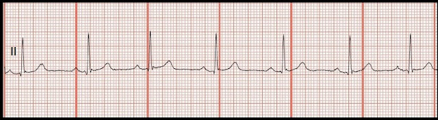
Practice EKG Rhythm Strips 218

Image
Identify the following rhythms. 1. 2. 3. 4. 5. Answers 1. Sinus arrhythmia The rhythm is irregular.   The rate is 70/min.   There are uniform, upright P waves before each QRS complex.  No ectopic beats are noted.  PR:  . 20 sec,  QRS:  .08 sec,  QT:  .38 sec.   Interpretation:  Sinus arrhythmia 2.  Sinus rhythm with a pause The rhythm is irregular.   The heart rate is 100/min.   The P waves are positive and precede each QRS complex.   There is a 1.08 second sinus pause that follows the 3rd complex.  No ectopic beats are noted.   PR:  .12 sec,  QRS:  .08 sec,  QT:  .32 sec.   Interpretation:   Sinus rhythm with a pause 3. Sinus bradycardia with sinus arrhythmia The rhythm is irregular.   The rate is 50/min.   P waves are present, upright, and associated with a QRS c...

Practice EKG Rhythm Strips 212

Image
Identify the following rhythms. 1. 2. 3. 4. 5. Answers 1. Junctional rhythm with bigeminal PVCs The rhythm is irregular with a rate of 60/min.  No P waves are seen.  There are bigeminal PVCs present.   PR:  ---,  QRS:  .08 sec,  QT:  .44 sec.   Interpretation:   Junctional rhythm with bigeminal PVCs 2. Normal sinus rhythm Wow, this strip looks really dirty.   I apologize.  Something must have changed in the contrast when it was uploaded. The rhythm is regular with a rate of 60/min.  There are P waves associated with each QRS complex.  The P waves are upright and uniform.  There is ST depression and T wave inversion seen in lead II and 1mm ST elevation seen in lead V1.  PR:  .18 sec,  QRS:  .08 sec,  QT:  .36 sec.  Interpretation:  Normal sinus rhythm 3. Normal sinus rhythm with PACs The rhythm is irregular. ...

Practice EKG Rhythm Strips 169

Image
Identify the following rhythms. 1. 2. 3. 4. 5. Answers 1. Junctional rhythm The rhythm is regular.   The rate is 60.   The P waves are absent.  No ectopic beats are noted. PR: ---,  QRS:  .12 sec,  QT:  .44 sec 2. Multifocal atrial tachycardia The rhythm is irregular due to the many PACs.   The rate is 140/min.   There are upright P waves present of differing morphology.   Some P waves are buried with the T wave of the previous complexes.  To be multifocal atrial tachycardia there needs to be at least three P waves of various morphology with a ventricular rate over 100/min.  PR---,  QRS:  .08,  QT:  .32 sec 3. Normal sinus rhythm with sinus pause The rhythm is irregular due to the pause.   The rate is 60/min.   The P waves are upright and are associated with a QRS complex.  There is...

Practice EKG Rhythm Strips 157

Image
Identify the following rhythms. 1. 2. 3. 4. 5. Answers 1. 1st degree block The rhythm is regular and the rate is 65/min.   There are uniform, upright P waves before each QRS complex.  However the PR interval is prolonged.   No ectopy is noted.   PR:  .32 sec,  QRS:  .08 sec,  QT:  .36 sec. 2. 2nd degree AV block type II The rhythm is regular.   The P-P rhythm is regular as well as the R-R interval.   The atrial rate is 120/min and the ventricular rate is calculated to be 34/min.  The P waves are upright and uniform.  However, there are two non-conducted P waves for each QRS complex.   Some P waves are buried in the T waves.    The PR interval is consistent.  PR:  .20 sec.   QRS:  .10 sec,  QT:  .44 sec.   3. 2nd degree AV block type I The rhythm ...