Posts

Showing posts with the label SVT

Sinus Rhythm Changing to SVT

Image
Sinus Rhythm Changing to SVT

Practice EKG Rhythm Strips 215

Image
Identify the following rhythms. 1. 2. 3. 4. 5. Answers 1. 2nd degree AV block type I The rhythm is irregular with a rate of 60/min.   There are upright P waves present.   There is progressive elongation of the PR interval over successive beats.   A non-conducted P wave follows the 4th complex.   No ectopic beats are present.   The QRS complex is narrow.   PR:  .28/.32/.36 sec.  QRS:  .08 sec,  QT:  .44 sec.   Interpretation:  2nd degree AV block type I 2. Atrial paced rhythm  The rhythm is regular with a rate of 75/min.   There are small upright P waves that follow the atrial pacer spike.  No ectopic beats are seen.  PR:  .28 sec.  QRS:  .08 sec,  QT:  .34 sec.   Interpretation:  Atrial paced rhythm 3. NSR with a pause followed by a junctional escape beat  The rhythm is irregular.   The...

Practice EKG Rhythm Strips 209

Image
Identify the following rhythms. 1. 2. 3. 4. 5. Answers 1. Atrial flutter with variable ventricular response The rhythm is irregular with a rate of 60/minute.   Multiple P waves or flutter waves are present in a 3:1 and 4:1 ratio.   No ectopic beats are seen.  PR:---,  QRS: .08 sec,  QT: .24 sec. Interpretation: Atrial flutter with variable ventricular response 2. Complete heart block The rhythm is regular with a rate of 38/min.   There are some small upright P waves seen but they are associated with the QRS complexes.   The atrial rate is 60/min.   No ectopic beats are seen.  PR:  ---,  QRS:  .16 sec,  QT:  .44 sec.   Interpretation:  Complete heart block 3. Normal sinus rhythm The rhythm is regular with a heart rate of 100/min.   The P waves are uniform, upri...

Practice EKG Rhythm Strips 208

Image
Identify the following rhythms. 1. 2. 3. 4. 5. Answers 1. Compete heart block The rhythm is regular with a ventricular rate of 36/min and an atrial rate of 83/min.   There is complete AV dissociation so the P waves do not have a corresponding QRS complex.   The P waves are uniform and upright suggesting a sinus origin.   The QRS complex is wide suggesting a ventricular escape mechanism that is stimulating the ventricles.   PR:  ---,  QRS:  .12 sec,  QT:  .52 sec. 2. Supraventricular tachycardia The rhythm is regular with a heart rate of 150/min.   The P waves are not readily seen.  The QRS complex is narrow which is indicative of a supraventricular rhythm.  The rhythm has a saw tooth appearance which resembles atrial flutter but until the rate is slowed down we can not be sure. Vagal maneuvers may provide some clues No ectopic beats are seen....

Practice EKG Rhythm Strips 196

Image
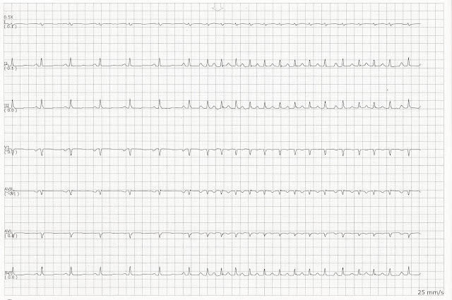
Identify the following rhythms. 1. 2. 3. 4. 5. Answers 1. Sinus bradycardia with 1st degree block The rhythm is regular with a rate of 65/min.   The P waves are uniform, positive, and precede the QRS complex.  However, the PR interval is prolonged.   No ectopic beats are present.   PR:  .38 sec,  QRS:  .10 sec,  QT:  .40 sec. 2. Sinus tachycardia The rhythm is regular with a heart rate of 136/min.   The P waves are small but they are upright and positive.   No ectopic beats are noted.   PR:  .12 sec,  QRS:  .08 sec,  QT:  .28 sec. 3. Sinus tachycardia with 1st degree block The rhythm is regular.   The heart rate is 100/min.   The P waves are buried in the T wave of the preceding complex.  You can see a P wave on the later half of the T wave of the 1st and 3rd complexes but less clearly on t...

Practice EKG Rhythm Strips 194

Image
Identify the following rhythms. 1. 2. 3. 4. 5. Answers 1. NSR with multifocal PVCs The rhythm is irregular due to the PVCs.   The heart rate is 70/min.   The P waves are wide, upright, and have a corresponding QRS complex.   The wide P waves suggests left atrial enlargement.   Multifocal PVCs are present.   PR:  .20 sec,  QRS:  .08 sec,  QT:  .40 sec. 2. Sinus arrhythmia The rhythm is irregular with a ventricular rate of 70/min.  The R-R interval between the 1st and 2nd complex is  0.8 sec and the R-R interval between the 5th and 6th complexes is 1.0 sec.  The P waves are upright and uniform with a fixed PR interval.   No ectopic beats are noted.   PR:  .16 sec,  QRS:  .08 sec,  QT:  .84 sec. 3. Sinus bradycardia The rhythm is regular with a heart rate of 47/min.   The P waves are positive and associated with a QRS...