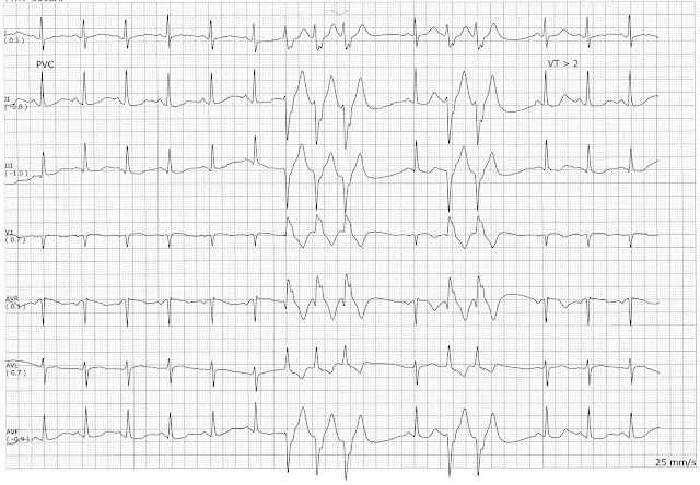
Identify the following rhythms. 1. 2. 3. 4. 5. Answers 1. Complete heart block The rhythm is regular with a ventricular rate of 38/min. There are small upright P waves present but they are not associated with a QRS complex. The QRS complexes are wide suggesting a ventricular origin. No ectopic beats are seen. PR: ---, QRS: .16 sec, QT: .40 sec. Interpretation: Complete heart block 2. Normal sinus rhythm The rhythm is regular. The rate is 75/min. There are upright P waves before each QRS complex. Lead II shows 3mm ST segment depression with an inverted T wave. No ectopic beats are seen. PR: .16 sec, QRS: .08 sec, QT: .36 sec. Interpretation: Normal sinus rhythm 3. Normal sinus rhythm with a multifocal couplet This strip shows an arterial blood pressure waveform in the 2nd lead. ...