Posts

Showing posts with the label 2nd degree block type I

Second degree Type I changing to second degree type II
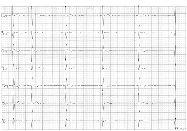
Image
Second degree Type I changing to second degree type II.  This patient switched back and forth between a type I block and a type II block.  

2nd Degree Heart Block Type I

Image
On this page you can see the progressive prolongation of the PR interval over successive beats.  The dropped beat is not seen in lead I but you can see it clearly in leads II and aVR.  A PVC interrupts the cycle of beats.  Instead of continuing in the cycle, the cycle of beats starts over after a compensatory pause.

Intermittent Type I Block

Image
This patient had an intermittent type I block.  He would have periods of sinus rhythm and then have periods of 2nd degree type I block.  The grouping on the type I block was not always consistent.  It may have been 3 beats in one group and 5 beats in another group.

Practice EKG Rhythm Strips 221

Image
Identify the following rhythms. 1. 2. 3. 4. 5. Answers 1. 2nd degree heart block type I The rhythm is irregular with a rate of 60/min.   The P waves are uniform and upright.   There is some splitting of the P wave which is often seen with biatrial enlargement.   There is progressive elongation of the PR interval over successive beats.   There is a dropped P wave after the 2nd QRS complex.   The QRS complexes are narrow.   PR:  .20/.24/..26/.30 sec.   QRS:  .08 sec,  QT:  .44 sec.   Interpretation:  2nd degree heart block type I 2. Accelerated junctional changing to sinus rhythm The rhythm is slightly irregular.   The heart rate is 83/min.   The P waves are negative or possibly biphasic in complexes 1 through 5 and then are positive in the remaining complexes.   The QRS complexes are narrow.  PR:  .10 sec,  QRS: ...

Practice EKG Rhythm Strips 215

Image
Identify the following rhythms. 1. 2. 3. 4. 5. Answers 1. 2nd degree AV block type I The rhythm is irregular with a rate of 60/min.   There are upright P waves present.   There is progressive elongation of the PR interval over successive beats.   A non-conducted P wave follows the 4th complex.   No ectopic beats are present.   The QRS complex is narrow.   PR:  .28/.32/.36 sec.  QRS:  .08 sec,  QT:  .44 sec.   Interpretation:  2nd degree AV block type I 2. Atrial paced rhythm  The rhythm is regular with a rate of 75/min.   There are small upright P waves that follow the atrial pacer spike.  No ectopic beats are seen.  PR:  .28 sec.  QRS:  .08 sec,  QT:  .34 sec.   Interpretation:  Atrial paced rhythm 3. NSR with a pause followed by a junctional escape beat  The rhythm is irregular.   The...

Practice EKG Rhythm Strips 204

Image
Identify the following rhythms. 1. 2. 3. 4. 5. Answers 1. Complete heart block The rhythm is regular with a ventricular rate of 35 and an atrial rate of 90/min.   The P waves are upright and uniform but are not associated with a QRS complex.   No ectopic beats are present.   The wide QRS complex suggests a ventricular escape mechanism.    PR:  ---,  QRS:  .12 sec.  QT:  .40 sec. 2. 2nd degree AV block with a PVC The rhythm is irregular with a heart rate of 40/min.   The P waves are upright and uniform.   There are P waves associated with some of the QRS complexes but there are also some dropped complexes.  On the conducted beats, the PR interval is fixed.  A single PVC is present.   PR:  .36 sec.   QRS:  12 sec,   QT:  .56 sec, 3. 1st degree heart block ...

Practice EKG Rhythm Strips 202

Image
Identify the following rhythms. 1. 2. 3. 4. 5. Answers 1. 2nd degree heart block type II The rhythm is irregular with a ventricular rate of 20/min and an atrial rate of  75/min.   The P waves are upright but not all of them are paired with a QRS complex.  No ectopic beats are noted.  The QRS complex is wide.   PR:  .20 sec,  QRS:  .12 sec,  QT:  .64 2. 2nd degree heart block type I The rhythm is irregular with a heart rate of 50/min.   The P waves are upright and uniform.    There is a nonconducted P wave after the 2nd complex.    The PR interval progressively lengthens over successive beats.   PR:  .20/.24/.32sec,  QRS:  .08 sec,  QT:  .40 sec 3. 3rd degree heart block The rhythm is regular with a ventricular rate of 30/min and an atrial rate of 100/min.   The P waves are upright and unifor...

Practice EKG Rhythm Strips 195

Image
Identify the following rhythms. 1. 2. 3. 4. 5. Answers 1. Complete heart block The rhythm is regular with a ventricular rate of 35/min and an atrial rate of 100/min.   The P waves and the QRS complexes are not associated.   The wide QRS complexes with a slow rate is indicative of  a ventricular escape rhythm.   PR:  ---,  QRS:  .16 sec,  QT:  .40 sec. 2. NSR with a multifocal couplet The rhythm is is irregular due to the PVCs.   The heart rate is 90/min.   The P waves are upright and appear before the QRS complexes.  A multifocal couplet is present.  The rSR pattern in the V1 lead suggests a RBBB.   There is 1mm ST depression in lead II.  PR:  .16 sec,  QRS:  .12 sec,  QT:  .36 sec. 3. NSR with trigeminal PACs The rhythm is irregular with frequent PACs.   The rate is 100/min.  ...